Gilded silver plates of the Great Shrine of st Maurice
Romain. Jeanneret (Abbaye de St-Maurice, Saint-Maurice, Valais, Switzerland)
 Credit ABSM, R.Jeanneret.
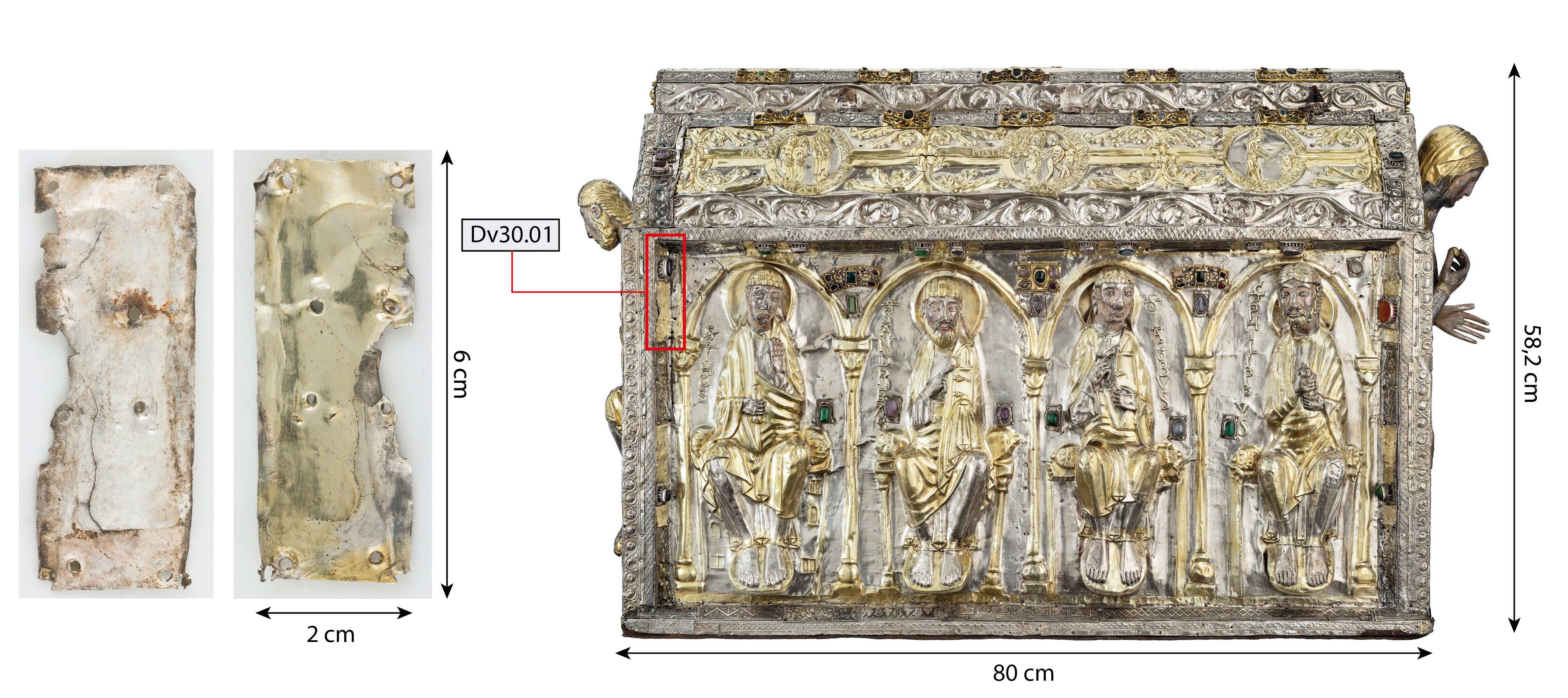
These two pieces are part of 44 gilded or partially gilded silver plates of the Great Shrine of saint Maurice with evidence of earlier decoration. These gilded plates are found re-used as backgrounds for set gems and filigree plates.
Religious goldsmithing
Northern continental Europe
Around 1160
High medieval times
Identified as re-used from a Romanesque relief dated around 1160
Indoor atmosphere
Abbaye de St-Maurice (Jeanneret Romain), Saint-Maurice, Valais
Abbaye de St-Maurice (Jeanneret Romain), Saint-Maurice, Valais
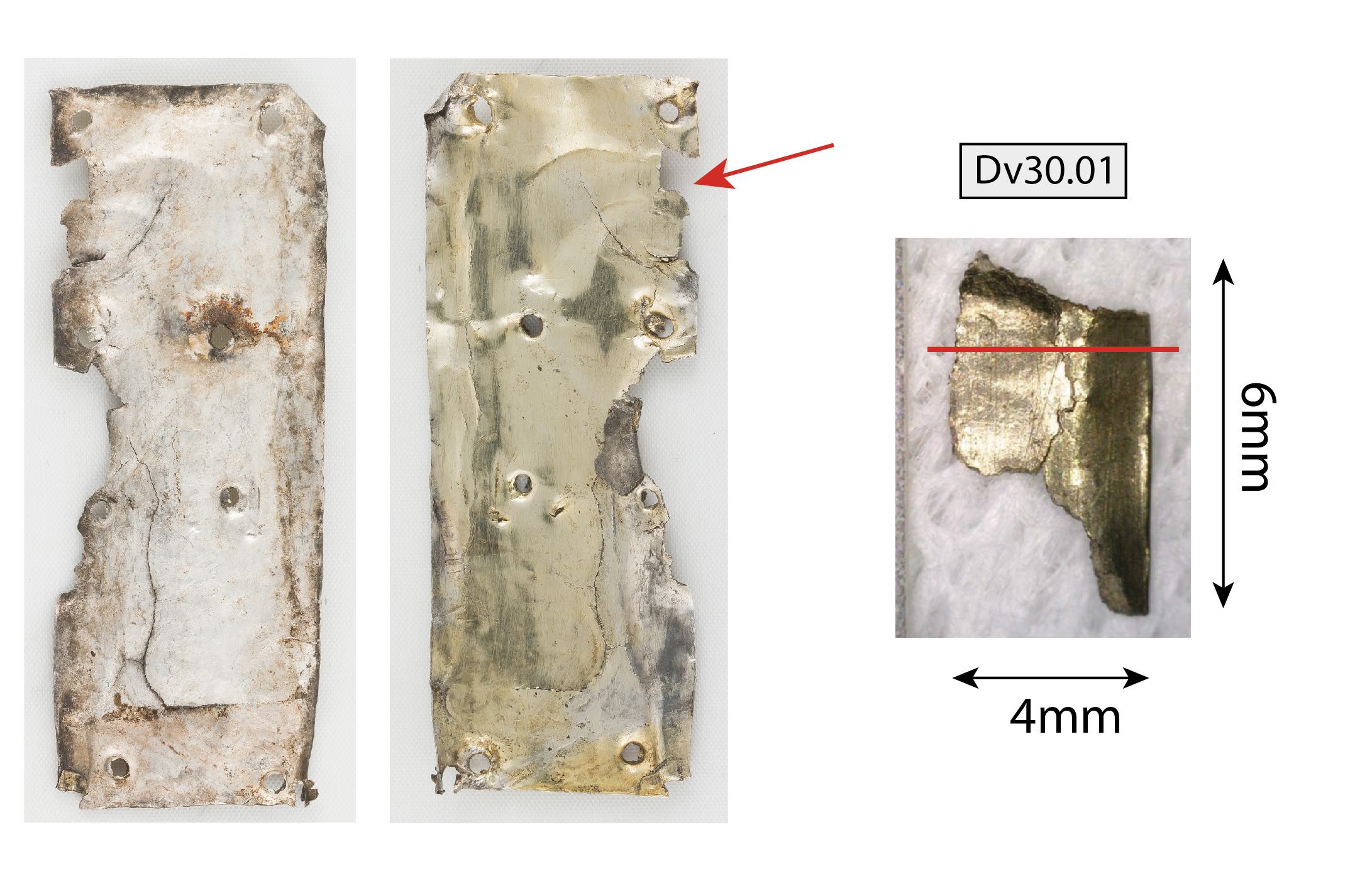
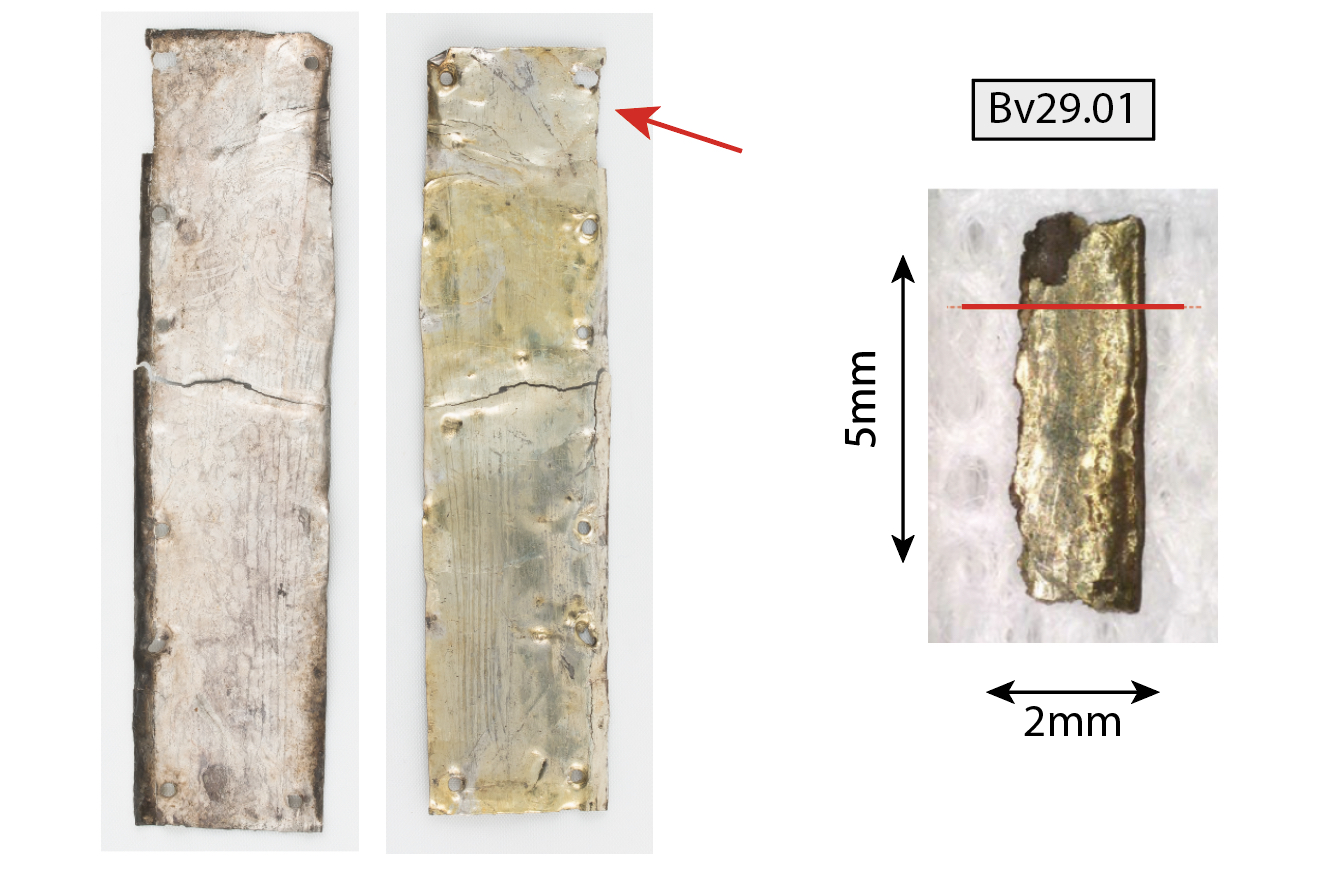
Inv.2_Bv29.01 & Dv30.01
The plates are a re-use of gilded reliefs, traces of which can still be found on the Great Shrine. The traces of the ancient decorations are similar to some of the ornaments still present, such as the mandorla of Christ, columns of the apostles, floral decorations, and ribbons flanking the medallions of the genesis present on the roof of the shrine.
For more information on the Great Shrine of saint Maurice see Nathania Girardin, 2015.
 Credit ABSM, R.Jeanneret.
None.
 Credit ABSM, R.Jeanneret.
 Credit HEI Arc, S.Ramseyer.
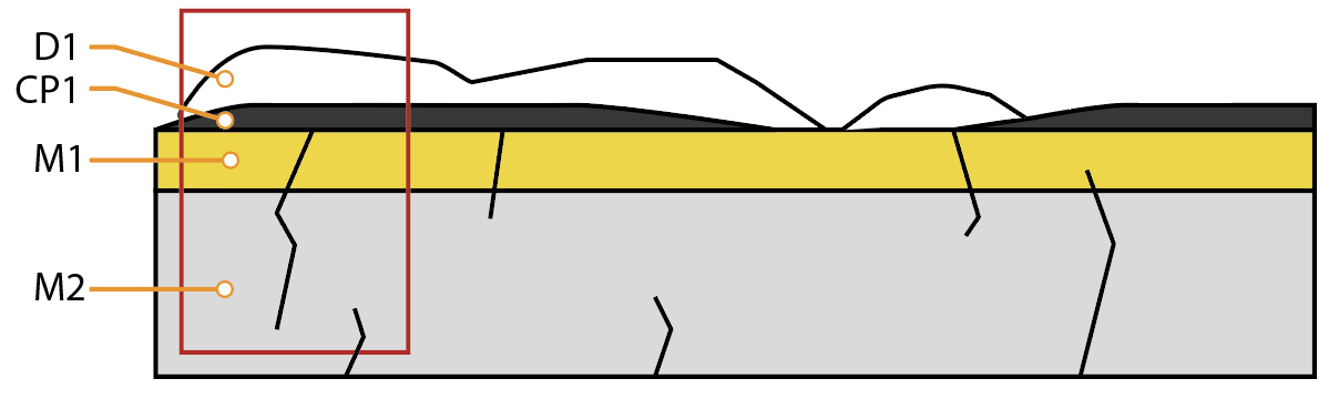
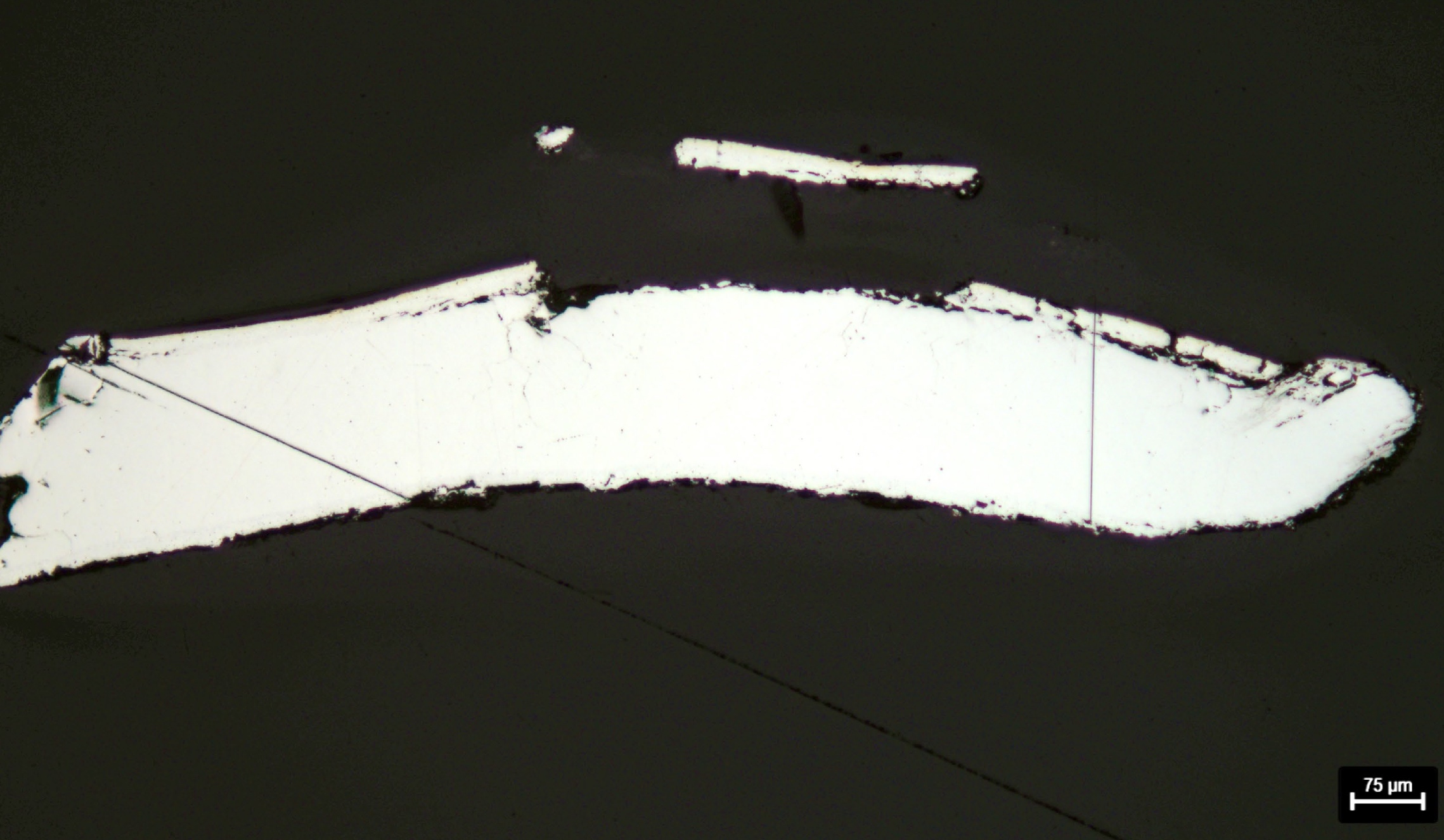
Samples of gilded plates fragments embedded (Figs. 7 & 8). These are transversal cuts as shown on Fig. 3 & 4.
Ag alloy
Hammered, annealed, repoussé, gilded and cold worked
Abbey of St Maurice, Saint-Maurice, Valais
Abbey of St Maurice, Saint-Maurice, Valais
18.09.2020
The plates Bv29.01 and Dv 30.01 are from the same decoration as the genesis reliefs. Intact examples of this can be found on the Great Shrine's roofs (Figs. 1 and 2). The plates have been cut out of the gilded areas and cold worked for reuse as gilded plates under gemstones and filigrees.
Analyses performed:
Non-invasive approach
- XRF with handheld portable X-ray fluorescence spectrometer (NITON XL3t 950 Air GOLDD+, Thermo Fischer®). Precious mode, acquisition time 60s.
Invasive approach (on samples)
- Optical microscopy: the sample is polished, then it is observed with a numerical microscope LEICA DMLM in bright field.
- Metallography: the polished sample is etched in an oxygenated ammoniacal solution (10mL NH3 25%+5mL H2O2 6%+10mL (NH4)2SO4 20%) and observed by optical microscopy in bright field.
- SEM-EDS: the sample is coated with a carbon layer and analyses are performed on a SEM-EDX JEOL equipped with a silicon-drift EDS Oxford detector with an accelerating voltage of 20 kV and probe current from a 1 to 10nA (to reveal the microstructure).
During a portable p-XFR analysis campaign, the ungilded sides of the plates were studied. The result of this analysis seems to indicate a silver alloy with about 1-2 wt%Cu. Trace elements such as gold and lead were also found.
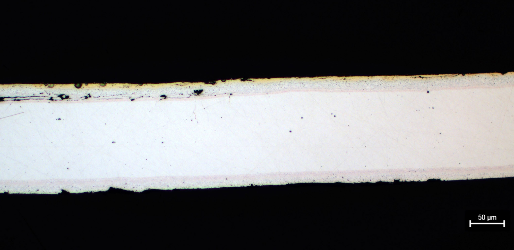
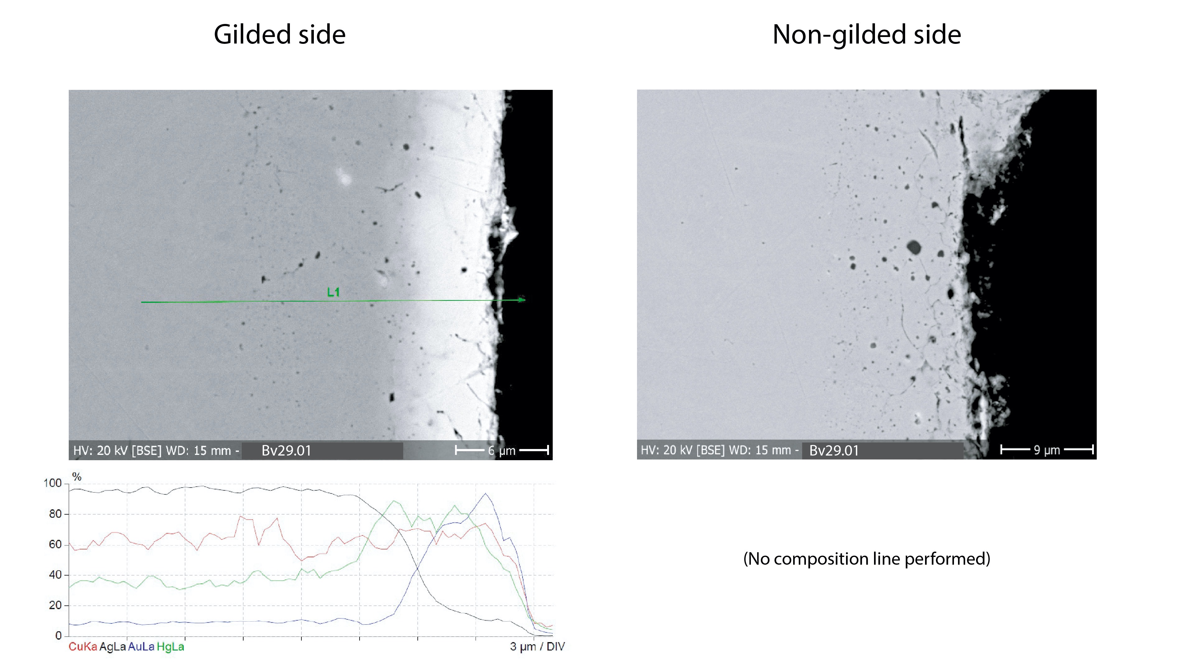
The metal of the plates is silver with a low percentage of copper (about 1-2wt%). The plates are gilded only on their visible side (Figs. 7 and 8). The gilding was studied in cross-section by EDS analysis using a composition line (Figs. 10 & 12). The results are similar for both plates and indicate a presence of mercury bound to both gold and silver and decreasing as we get away from the interface. These results are consistent with what has been studied by Kilian Anhauser on mercury gilding samples reproduced in the laboratory (Anhauser, 1999) and on medieval gilded silver (Schweizer & Degli Agosti, 2007).
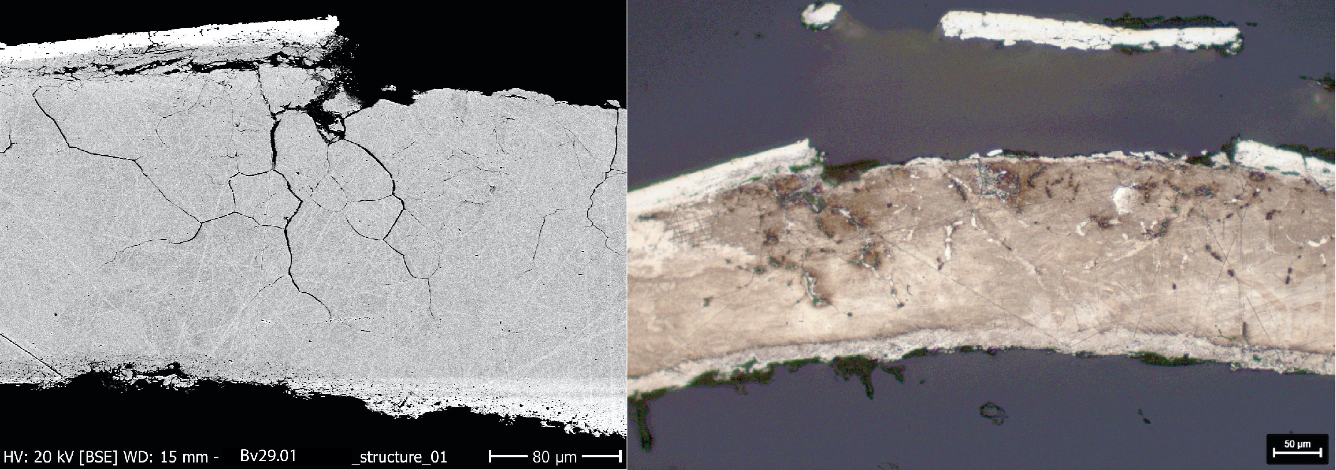
SEM images (Figs. 10 and 12) show that the metal surface of the underlying metal is slightly porous. This suggests a surface enrichment due to the repoussé work and successive annealing. This is reinforced by the etching of the two samples (Figs 9 & 11) indicating a very different reaction between the first 25 microns and the core of the alloys. Despite these differences, the composition line (Figs 10 & 12) analysis shows no variation in copper content.
Under the Dv30.01 gilding, there is a pink layer about 10um thick visible on the micrograph (Fig.8). While this may appear to be a highly enriched copper layer, it is not confirmed by composition line analysis. This layer is also found on the non-gilded side, indicating that it is maybe related to the gilding process. This is not clearly visible on plate Bv29.01.
The grain structure of the silver core (large polygonal grains, which may indicate recrystallization of the material(s)) is only visible in the SEM, BSE mode, and by increasing the beam intensity to 10nA (Figs. 9 and 11).
 Credit HEI Arc, S.Ramseyer & HE-Arc CR, C.Degrigny.
 Credit HEI Arc, S.Ramseyer
 Credit HEI Arc, S.Ramseyer & HE-Arc CR, C.Degrigny.
Recrystallized structure with large grains
Ag
Cu, Au, Hg
None.
The gilded metal is covered with a thin layer of brown to black tarnish on which deposits of cleaning products are detected. This tarnish on top of the gilding has been analyzed using Linear Sweep Voltammetry in the cathodic domain which showed a Ag2S reduction peak. Other works on tarnished gilded silver have shown the same results (Jeanneret & al. 2016).
The majority of cracks are due to cold work. It is possible that this phenomenon was amplified and/or followed by intergranular corrosion. Nevertheless, the mercury-enriched interface seems to have suffered the most from cold working leading to loss of local adhesion of the gilding. This could be explained by differences in the ductility of the different metal layers. Some of the cracks reach the center of the metal (Figs. 9 & 11).
On the reverse side of the plate, protected from the environment, only the edge is tarnished.
Passive
Silver tarnishing
None.
The observation in cross-section does not allow the identification of the surface deposits observed under the binocular microscope. This is due to the fact that the observations of the object are made before treatment while the fragment analyzed in cross-section has been treated (degreasing and reduction of tarnish). An intergranular corrosion may also have developed and increased the cracking.
The pinkish stratum (M3) and porous layer (M2) are clearly visible on the micrograph (Fig. 8) but compositional line analysis did not demonstrate any difference from the metal.
The analysis of the cross-section reveals the diffusion of mercury between the gilding and the silver, which is of course not visible under the binocular microscope.
The metal is a gilded silver alloy with a low copper content (about 1-2wt% Cu). As suspected, by naked-eye observation, it consists of a mercury gilding. The EDS analysis in line of composition shows well the presence of mercury at the interface between silver and gold and its diffusion in both metals.
Its corrosion layers are typical of gilded silver with a very thin tarnish that has not been analyzed but is considered to be a mixture of silver sulfide and chloride. We did not observe any copper corrosion on the surface during voltammetric measurements. As with other silver gilded pieces treated by electrolytic reduction (Jeanneret, R. 2016), it seems that the copper does not diffuse through the gilding, unlike silver.
The metallographic study shows a microstructure that is difficult to interpret as indicated below. Etching indicates a difference in reaction between the surface layer of about 30μm and the core of the silver alloy. This suggests a surface enrichment prior to gilding since the gold layer appears on top. The lack of difference in copper percentage shown by the EDS line of composition may be due to the original low content of copper within the alloy.
The several cracks on the two plates are the result of metal strain after cold work. This is the result of flattening the original repoussé reliefs to re-use its gilded surfaces as background plates for gems and filigree of the Great Shrine of saint Maurice. An intergranular corrosion may also have developed and increased the cracking.
References on object and sample
1. Girardin, N. (2015). La châsse de saint Maurice. In: Mariaux, P.A dir., L'abbaye de Saint-Maurice d'Agaune 515-2015. Volume 2 - Le trésor. Ed. Infolio. Gollion, 73-85.
References on analytic methods and interpretation
2. Anheuser, K. (1999). Im Feuer vergoldet. Geschichte und Technik der Feuervergoldung und der Amalgamversilberung. AdR Schriftenreihe zur Restaurierung und Grabungstechnik Band 4. Stuttgart.
3.Jeanneret, R., Degrigny, C., Fontaine, C. and Tarchini, A. (2016). Using the Pleco: Electrolytic Treatment of Metal Components on Artefacts. Conference: METAL 2016, proceedings of the ICOM-CC Metal WG interim meeting. New Dehli.
4.Schweizer, F. & Degli Agosti, M. (2007). Plaques en argent repoussé et ornements de moulures. In Schweizer, F. & Witschard, D. La Châsse des enfants de saint Sigismond. Ed. Somogy, 153-179.
5.Wanhill, R. (2013) Stress corrosion cracking in ancient silver, Studies in Conservation, 58:1, 41-49, DOI: 10.1179/2047058412Y.0000000037.