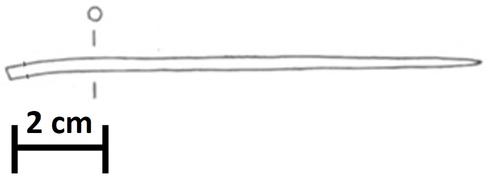
Pin or needle fragment HR-3031
Marianne. Senn (EMPA, Dübendorf, Zurich, Switzerland) & Christian. Degrigny (HE-Arc CR, Neuchâtel, Neuchâtel, Switzerland) & Naima. Gutknecht (HE-Arc CR, Neuchâtel, Neuchâtel, Switzerland) & Rémy. Léopold (HE-Arc CR, Neuchâtel, Neuchâtel, Switzerland)
 Credit HE-Arc CR, L.Rémy.
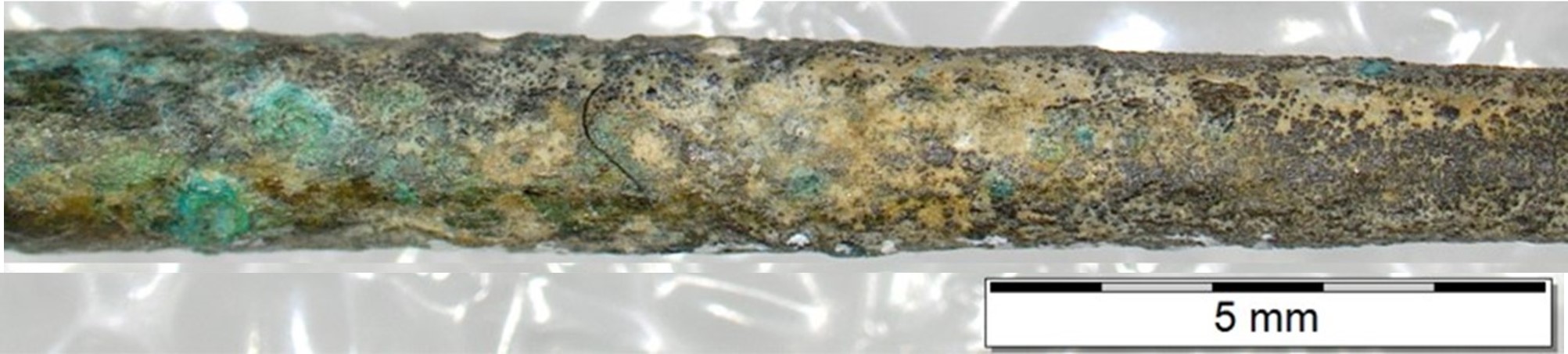
Pin or needle fragment (Fig. 1) with dark grey to dark green corrosion products (Figs. 2 & 3). Dimensions: L = 9cm; Ø = 2.5-2.9mm; WT = 3.6g.
Jewellery
Hauterive - Champréveyres, Neuchâtel, Neuchâtel, Switzerland
Excavation 1983-1985, object from layer 1 (containing material from the Bronze Age until the 20th century)
Late Bronze Age
Hallstatt A2/B
Lake
Laténium, Neuchâtel, Neuchâtel
Laténium, Neuchâtel, Neuchâtel
Hr 3031
Not conserved
Considered to be a land patina by Schweizer (1994). The object was sampled in 1987 for analysis. Documentation of the strata in binocular mode on the remaining fragment of the object was performed in 2022.
 Credit HE-Arc CR, L.Rémy.
 Credit Laténium, C.Cevey.
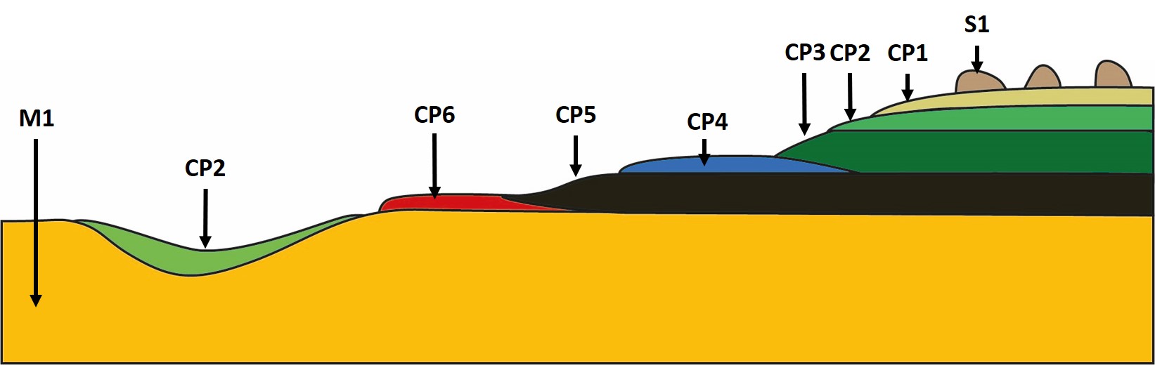
The schematic representation below gives an overview of the corrosion structure encountered on the pin from a first visual macroscopic observation.
| Strata | Type of stratum | Principal characteristics |
| S1 | Sediments | Nodule, light brown, thin, scattered, non compact, friable, very soft |
| CP1 | Corrosion product | Layer, light brown, thin, discontinuous, compact, powdery, soft |
| CP2 | Corrosion product | Layer, light green, thin, discontinuous, compact, powdery, soft |
| CP3 | Corrosion product | Layer, dark green, thin, discontinuous, compact, tough, soft |
| CP4 | Corrosion product | Layer, blue, thin, discontinuous, compact, tough, soft |
| CP5 | Corrosion product | Layer, black, thin, discontinuous, compact, tough, soft |
| CP6 | Corrosion product | Layer, dark red, thin, scattered, non compact, friable, soft |
| M1 | Metal | Dark yellow, thick, compact, soft |
Table 1: Description of the principal characteristics of the strata as observed under binocular and described according to Bertholon's method.
 Credit HE-Arc CR, L.Rémy.
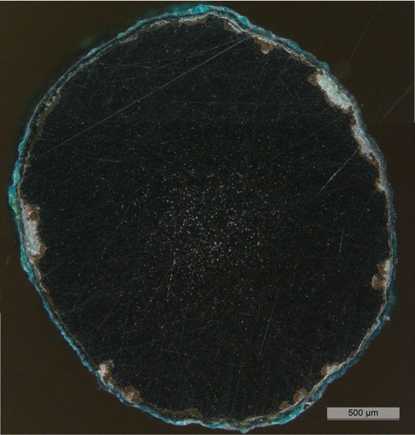
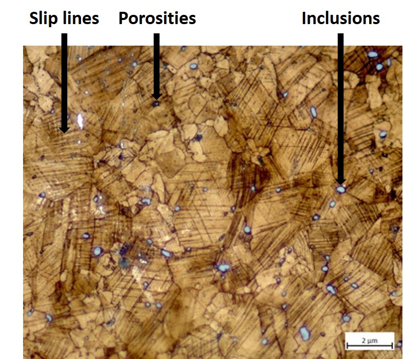
The cross-section is circular and is a complete section through the pin (Fig. 9). The surface is completely covered with a rather thin corrosion layer of irregular thickness (Fig. 10). At the center of the cross-section, the metal seems to have more porosities and/or inclusions.
Tin Bronze
Cold worked with partial annealing
MAH 87-195
Musées d'art et d'histoire, Genève, Geneva
Musées d'art et d'histoire, Genève, Geneva
1987, metallography and corrosion characterisation
This sample is mentioned in Schweizer, 1994.
Analyses performed:
Non-invasive approach
XRF with handheld portable X-ray fluorescence spectrometer (NITON XL5). General Metal mode, acquisition time 60s (filters: Li20/Lo20/M20).
Invasive approach (on the sample)
Metallography (etched with ferric chloride reagent), Vickers hardness testing, ICP-OES (conditions provided in the About tab of the MiCorr application), SEM/EDS (20 keV, Microcity), XRD.
XRF analysis of the pin was carried out on the surface of a representative area next to the cross-section. All strata (soil, corrosion products, and metal) are analyzed at the same time.
The metal is presumably a tin bronze alloy. The other elements detected are: Si, S, Al, Fe, Pb, Sb, As, Ag, Ni, P, Zn.
|
Elements (mass %) |
Cu |
Sn |
Si |
S |
Al |
Fe |
Pb |
Sb |
As |
Ag |
Ni |
P |
Zn |
|
|||||||||||||
|
|
% |
+\- 2σ |
% |
+\- 2σ |
% |
+\- 2σ |
% |
+\- 2σ |
% |
+\- 2σ |
% |
+\- 2σ |
% |
+\- 2σ |
% |
+\- 2σ |
% |
+\- 2σ |
% |
+\- 2σ |
% |
+\- 2σ |
% |
+\- 2σ |
% |
+\- 2σ |
Total |
|
1 |
75.5 |
0.2 |
9.5 |
0.05 |
5.0 |
0.1 |
2.0 |
0.04 |
2.0 |
0.2 |
1.5 |
0.03 |
1.0 |
0.02 |
1.0 |
0.02 |
0.7 |
0.03 |
0.4 |
0.01 |
0.2 |
0.02 |
0.1 |
0.01 |
0.1 |
0.01 |
99.0 |
Table 2: Chemical composition of the surface of the pin at one representative area shown in Fig. 4. Method of analysis: XRF, UR-Arc CR.
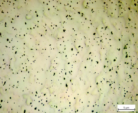
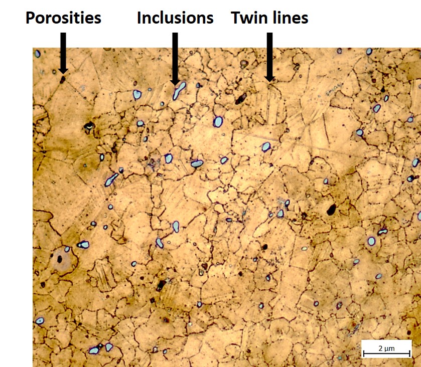
As suggested by XRF analysis, the remaining metal is a tin bronze with some Sb (Table 3), porosities and small copper sulphide inclusions (Table 4). The etched structure of the tin bronze shows two zones (Fig. 12): more than one half of the section is constituted of re-crystallised and angular grains, some of them with twins (Fig. 13), while the other half shows the presence of strain or slip lines in the grains (Fig. 14). They indicate a final cold working without annealing. Copper sulphide inclusions are found both at the grain boundaries and inside the grains (Figs. 13 and 14). The average hardness of the metal is about HV1 120.
| Elements | Cu | Sn | Sb | Ni | As | Pb | Ag | Co | Zn | Fe |
|---|---|---|---|---|---|---|---|---|---|---|
| mass% | 91.29 | 5.65 | 1.00 | 0.69 | 0.55 | 0.51 | 0.22 | 0.06 | 0.01 | 0.02 |
Table 3: Chemical composition of the metal. Method of analysis: ICP-OES, Laboratory of Analytical Chemistry, Empa.
| Elements | S | Cu | Total |
|---|---|---|---|
| mass% | 21 | 85 | 106 |
Table 4: Chemical composition of grey inclusions. Method of analysis: SEM/EDS, Laboratory of Analytical Chemistry, Empa.
 Credit HE-Arc CR, L.Rémy.
 Credit HE-Arc CR, L.Rémy.
 Credit HE-Arc CR, L.Rémy.
 Credit HE-Arc CR, L.Rémy.
Polygonal and twinned grains (one half of the cross-section) & slip lines (second half)
Cu
Ni, As, Ag, Sn, Sb, Pb
Schweizer (1994) indicates that the copper-tin alloys similar to the one of the pin have minor constituents that were certainly not added intentionally. Furthermore, he mentions that there is no systematic composition difference between bronzes with a lake patina and those with a land patina.
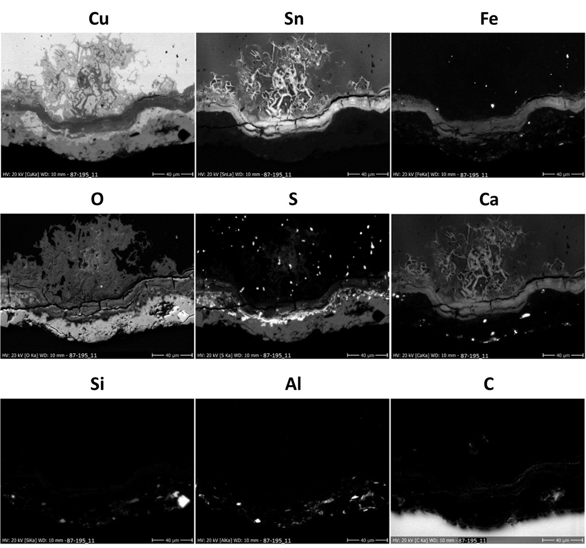
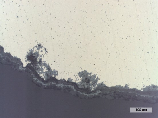
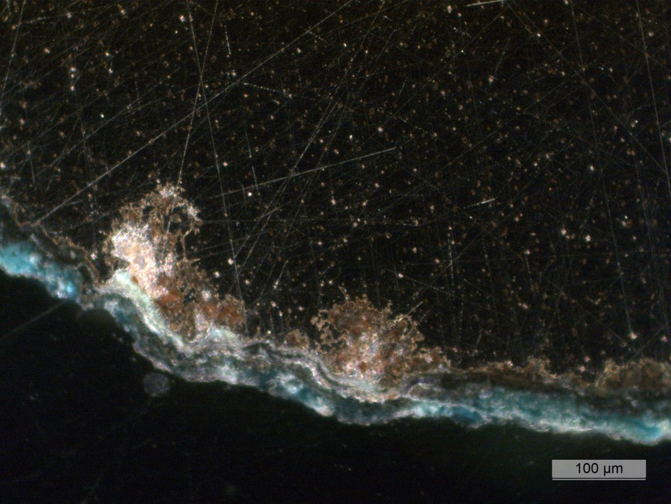
The corrosion layer has an average thickness of about 50µm (Fig. 15). The observation of the sample in cross-section (Figs. 15 and 18) shows the presence of a succession of layers (Figs. 17 and 18): S1 sediment at the top surface, discontinuous layer, black in bright field and light yellow in dark field; CP1 medium continuous layer, extra dark grey in bright field, dark turquoise in dark field; CP2 thin continuous layer, extra light grey in bright field, black in dark field; CP3 thin continuous layer, dark grey in bright field, extra light grey in dark field; CP4 thin continuous layer, extra dark grey in bright field, black in dark field; CP5 thin discontinuous layer, black in bright field, light green in dark field; CP6 medium continuous layer, light grey in bright field, dark orange in dark field; CM1 thin continuous layer;
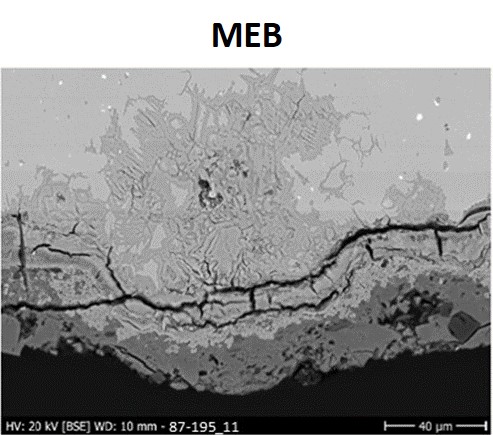
The EDX elemental mapping (Fig. 20) of SEM image (Fig. 19) of the visually identified strata in cross-section shows the following chemical distribution :
S1 has Al, Ca, Si, and O elements;
CP1 stratum contains Cu, O and S;
CP2 stratum contains Cu, S and Fe;
CP3 stratum contains Sn, O, Fe and S;
CP4 stratum contains Cu, Sn, O, Fe and S;
CP5 stratum contains Cu, Sn, O, Fe and S;
CP6 stratum contains Cu, Sn, O and S.
 Credit HE-Arc CR, L.Rémy.
 Credit HE-Arc CR, L.Rémy.
 Credit HE-Arc CR, L.Rémy.
 Credit HE-Arc CR, L.Rémy.
 Credit HEI Arc, S.Ramseyer.
Multiform - pitting
Type II (Robbiola)
None.
Sediment (S1) in binocular mode is documented as sediment (S1) in cross-section mode.
CP1, CP2, CP3 and CP4 in binocular mode are differentiated by their color. They match CP1 in cross-section mode.
CP5 in binocular mode matches CP2, CP3 and CP4 in cross-section mode.
CP6 in binocular mode seems to match CP5, CP6 and CM1 in cross-section mode.
On cross-section, it was possible to describe and analyze the microstructure of the metal.
The pin is made from a tin bronze and has been cold worked and partially annealed. Due to the presence in the corrosion layer of an outer malachite layer (to confirm), the corrosion was described as terrestrial by Schweizer (Schweizer 1994). The elemental chemical distribution of the corrosion layer shows a more complex situation: as expected for an object buried in a terrestrial site, a typical enrichment of Sn is observed in the inner and intermediate layers covering the remaining metal surface. However, it is combined with Fe and O. Cu, S and O are found on top layers. According to Schweizer, these layers were formed in anaerobic conditions and developed later on into malachite in an aerated soil through partial dehydration (Schweizer 1994, Schwartz 1934). The limit of the original surface most probably lies between the Sn-rich inner layer and the Fe/Cu and S-rich outer layers. We refer to type 1 corrosion after Robbiola et al. 1998.
This object was first sampled in 1987. Thanks to an extensive documentation on the cross-section and comparison with similar objects (see references), Schweizer defines a "land patina" typology on this object. In fact it is close to Tang fragment of a knife Hr 6246.
References on object and sample
Object files in MiCorr
1. MiCorr_Pin HR-18152
2. MiCorr_Tang fragment of a knife HR-6567
3. MiCorr_Tang fragment of a knife HR-6246
4. MiCorr_Pin HR-17773
5. MiCorr_Pin HR-3071
6. MiCorr_Pin HR-18603
7. MiCorr_Pin HR-3389
References object
8. Rychner-Faraggi A-M. (1993) Hauterive – Champréveyres 9. Métal et parure au Bronze final. Archéologie neuchâteloise, 17 (Neuchâtel), planche 74.11.
References sample
9. Empa Report 137 695/1991, P.O. Boll.
10. Rapport d'examen, Laboratoire Musées d'art et d'Histoire, Geneva GE (1987), 87-194 à 197.
11. Schwartz, G.M. (1934) Paragenesis of oxidised ores of copper, Economic Geology, 29, 55-75.
12. Schweizer, F. (1994) Bronze objects from Lake sites: from patina to bibliography. In: Ancient and historic metals, conservation and scientific resarch (eds. Scott, D.A., Podany, J. and Considine B.B.), The Getty Conservation Institute, 33-50.
References on analytic methods and interpretation
13. Interpretation of orange corrosion products, see: Piccardo P., Mille B., Robbiola L. (2007) Tin and copper oxide in corroded archaeological bronzes, In: Corrosion of metallic heritage artefacts, European Federation of Corrosion Publication n°48, ed. Dillmann et al., 239-262.
14. Robbiola, L., Blengino, J-M., Fiaud, C. (1998) Morphology and mechanisms of formation of natural patinas on archaeological Cu-Sn alloys, Corrosion Science, 40, 12, 2083-2111.