Sickle AUV-322
Rémy. Léopold (HE-Arc CR, Neuchâtel, Neuchâtel, Switzerland)
 Credit HE-Arc CR, L.Rémy.
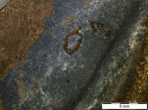
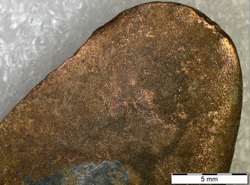
Sickle with a groove on the external front side and brown-yellow/dark-grey corrosion products (Figs. 1-3). Dimensions: L = 12.0cm; Ømax. = 3.1cm; WT = 91.0g.
Tool
Hauterive - Champréveyres, Neuchâtel, Neuchâtel, Switzerland
Excavation in 1971
Late Bronze Age
Lake
Laténium, Neuchâtel, Neuchâtel
Laténium, Neuchâtel, Neuchâtel
AUV 322
No conservation data available, but a coating and inventory number is visible on the surface.
The object was documented in 1987 by Valentin Rychner. Documentation of the strata in binocular mode on the object was performed in 2022.
 Credit HE-Arc CR, L.Rémy.
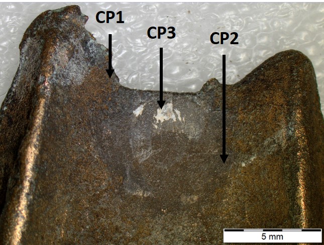
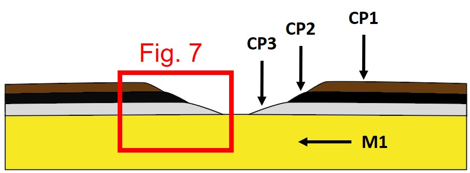
The schematic representation below gives an overview of the corrosion structure encountered on the sickle from a first visual macroscopic observation.
| Stratum | Type of stratum | Principal characteristics |
| CP1 | Corrosion product | Brown, matte, thin, discontinuous, compact, powdery, very soft |
| CP2 | Corrosion product | Black, matte, thin, discontinuous, compact, powdery, very soft |
| CP3 | Corrosion product | Extra light grey, matte, thin, discontinuous, compact, powdery, hard |
| M1 | Metal | Yellow, thick, metallic, continuous, compact, tough, very hard |
Table 1: Description of the principal characteristics of the strata as observed under binocular and described according to Bertholon's method.
Bronze
None
None
None
None.
Analyses performed:
Non-invasive approach
XRF with handled portable X-ray fluorescence spectrometer (NITON XL5), General Metal mode, acquisition time 60s (filters: Li20/Lo20/M20).
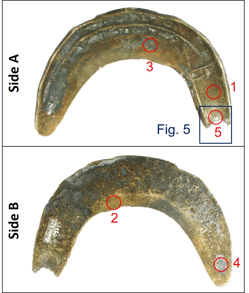
XRF analyses of the sickle were carried out on five representative areas (Fig. 4). Points 1 and 2 were done in the brown corrosion layer of each side (CP1), points 3 and 4 on the black corrosion layer of each side (CP2), and point 5 in the extra light grey of side A (CP3). All strata (soil, corrosion products, and metal) are analyzed at the same time.
The metal is presumably a tin bronze alloy with some As, Pb and Sb. The other elements detected are : S, Al, Si, Ni, Ag, Bi, P.
Results of points 1, 2 and 5 are very similar and give concentrations close to those of the remaining metal surface.
Results of points 3 and 4 are similar but different from those of points 1, 2 and 5: they indicate an enrichment in S and the depletion in Cu.
| Elements (mass %) | Cu | Sn | S | As | Pb | Al | Sb | Si | Ni | Ag | Bi | P | Fe |
||||||||||||||
| % | +/-2σ | % | +/-2σ | % | +/-2σ | % | +/-2σ | % | +/-2σ | % | +/-2σ | % | +/-2σ | % | +/-2σ | % | +/-2σ | % | +/-2σ | % | +/-2σ | % | +/-2σ | % | +/-2σ | TOTAL | |
| 1 | 83.1 | 0.1 | 7.3 | 0.04 | 2.0 | 0.02 | 2.0 | 0.04 | 1.6 | 0.03 | 1.0 | 0.08 | 0.9 | 0.02 | 0.8 | 0.03 | 0.5 | 0.02 | 0.4 | 0.01 | 0.1 | 0.01 | 0.1 | 0.01 | 0.1 | 0.01 | 99.7 |
| 2 | 83.2 | 0.1 | 7.8 | 0.04 | 2.6 | 0.02 | 1.4 | 0.03 | 1.0 | 0.02 | 0.8 | 0.07 | 0.9 | 0.02 | 1.1 | 0.03 | 0.4 | 0.01 | 0.3 | 0.01 | 0.1 | 0.01 | 0.1 | 0.01 | 0.1 | 0.01 | 99.8 |
| 3 | 72.0 | 0.15 | 7.3 | 0.04 | 11.4 | 0.06 | 1.3 | 0.03 | 1.1 | 0.02 | 1.5 | 0.15 | 0.9 | 0.02 | 3.4 | 0.08 | 0.5 | 0.02 | 0.3 | 0.01 | 0.1 | 0.01 | 0.1 | 0.02 | 0.2 | 0.01 | 100.1 |
| 4 | 70.8 | 0.15 | 8.6 | 0.05 | 10.6 | 0.07 | 1.7 | 0.04 | 2.3 | 0.03 | 0.8 | 0.15 | 0.8 | 0.02 | 2.7 | 0.08 | 0.4 | 0.01 | 0.3 | 0.01 | 0.1 | 0.01 | 0.4 | 0.03 | 0.3 | 0.02 | 99.8 |
| 5 | 80.7 | 0.2 | 7.6 | 0.05 | 1.6 | 0.04 | 1.7 | 0.04 | 1.6 | 0.03 | 1.5 | 0.02 | 0.9 | 0.03 | 2.0 | 0.08 | 0.4 | 0.02 | 0.4 | 0.01 | 0.1 | 0.01 | 0.1 | 0.02 | 1.1 | 0.02 | 99.7 |
Table 2: Chemical composition of the surface of the pin at five representative points shown in Fig. 4, Method of analysis: XRF, UR-Arc CR.
None.
None
Cu
Sn
Rychner (1987) indicates that the metal of the object is bronze.
The appearance of CP1 and CP2 and their composition (Cu, S) seem to indicate that they might be chalcocite or djurleite.
None
None
According to Rychner (1987), the dark corrosion layer (CP1) was previously analysed by XRD, it was identified as a mix of chalcocite (Cu2S) and djurleite (Cu1.93S).
The corrosion structure has only been documented in binocular mode (Fig. 7).
The sickle is made from a tin bronze. The XRF analysis shows that the black corrosion layer CP2 has higher %S and lower %Cu, it would indicate the presence of black copper sulfide such as chalcocite (Cu2S) and djurleite (Cu1.93S), as described by Rychner (1987).
References on object and sample
Object files in MiCorr
1. MiCorr_Sickle Auv-310
2. MiCorr_Sickle Auv-313
References object