Double-sided oil painting on iron plate with hangers
Zala. Uršič (, Ljubljana, Ljubljana, Slovenia) & Nataša. Nemeček (National museum of Slovenia, Ljubljana, Ljubljana, Slovenia)
 Credit National museum of Slovenia_NMS, Z.Ursic.
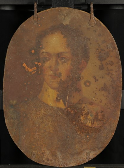
The painting is painted on both sides using the technique of oil on iron plate. It has two iron hooks on the upper edge and an oval shape. It measures 55.5 × 41 cm without hooks or 60.5 × 41 cm with hooks.
Painting
Unknown
After 1945
Second half of 19th century
Outdoor to indoor atmosphere
National museum of Slovenia (NMS), Ljubljana
National museum of Slovenia (NMS), Ljubljana
N 39363
The painting is currently in process of conservation and restoration.
On both sides of the painting there is a thick layer of dirt (made up of corrosion products, dust, soot and pollutants) and a yellowed thin layer of coating present (presumably oil-resin based). Paint layer on both sides is damaged (missing paint layer to the support due to flaking and corrosion). Areas where iron is exposed are corroded; brown, red and black corrosion products are visible. Orange corrosion products are also locally present on top of the paint layer.
 Credit National museum of Slovenia_NMS, Z.Ursic.
 Credit National museum of Slovenia_NMS, Z.Ursic.
 Credit National museum of Slovenia_NMS, Z.Ursic.
 Credit National museum of Slovenia_NMS, Z.Ursic.
 Credit National museum of Slovenia_NMS, Z.Ursic.
None.
 Credit National museum of Slovenia_NMS, Z.Ursic.
 Credit Restavratorski center_RC, K. Kavkler, Z.Ursic.
 Credit Restavratorski center_RC, K. Kavkler, Z.Ursic.
 Credit Restavratorski center_RC, K. Kavkler, Z.Ursic.
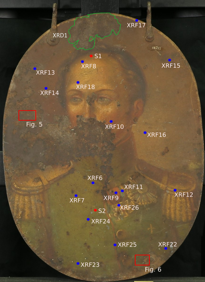
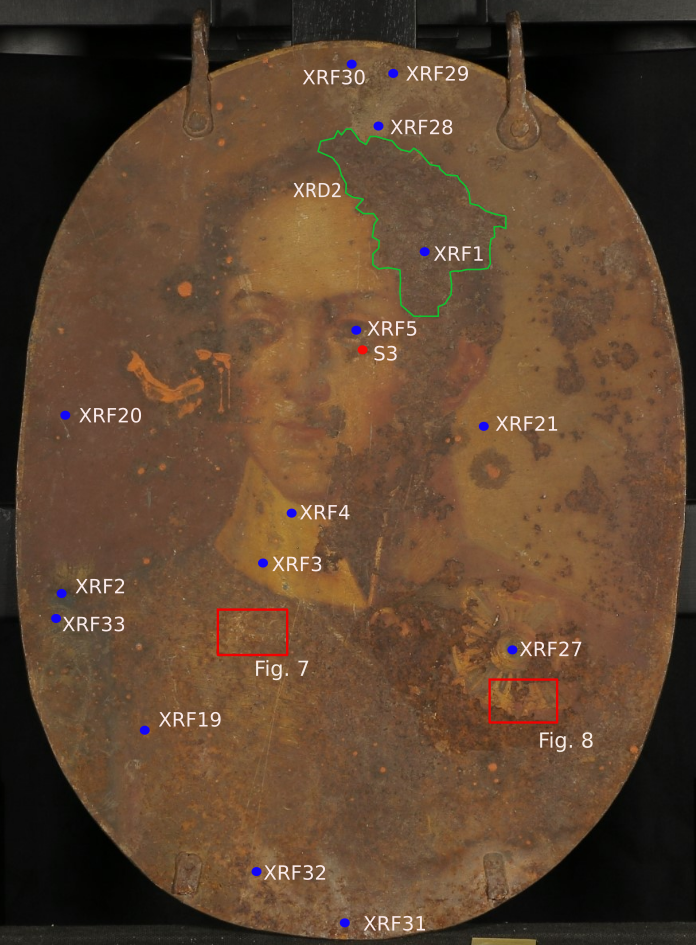
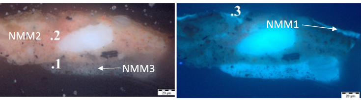
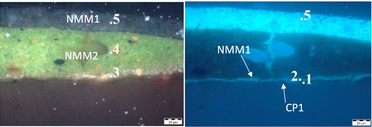
Samples (flakes) of paint layer were taken with a scalpel on various areas on both sides of the painting (see Fig. 3-4). Samples were embedded in polystyrene resin and grinded down to get a visible cross-section of paint layer.
Two 0.5 g samples for XRD analysis of corrosion products were taken from corroded area on each side of the painting (see Fig. 3-4). Samples were taken mechanically with a scalpel and were in powder form. Collected samples in powder form were also used for additional SEM-EDS analysis.
Fe Alloy
Unknown
1-2 (XRD, SEM-EDS), 1-3 (paint statigraphy).
Faculty of Natural Sciences and Engineering, Ljubljana
Faculty of Natural Sciences and Engineering, Ljubljana
March 2022 (statigraphy of paint layers), January- April 2023 (XRD and SEM-EDS identification of corrosion products).
Microscopic analysis of paint statigraphy showed that ground layer is very thin and has blue-gray metallic appearance. Additional XRF analysis of ground layer showed that it consists mainly of lead oxides, possibly with addition of zinc. The colour of ground layer is very unusual (metallic blue) and has not been explained yet.
Paint layer consists of different inorganic pigments with linseed oil as a binder. With additional XRF analysis we identified various pigments, such as lead white, emerald green, Naples yellow, chrome red, vermillion, various iron oxide pigments... With identification of those pigments and their historic use we were able to date the painting to the second half of 19th century.
The protective varnish layer consists of thin layer of organic yellowed material, most likely drying oil (linseed oil) or some type of resin.
Analyses performed:
Non invasive approach:
-XRF with handheld handheld portable ED X-ray fluorescence spectrometer (Hitachi X-MET 8000), calibration Alloy LE FP, 2 scans for each measurement; at 8 kV and 40 kV.
Invasive approach:
-XRD (no info for equipment yet)
-SEM-EDS
Microscopic analysis of paint statigraphy: sample was embedded in polystyrene resin and polished to get a cross-section, then observed with Olympus BX-60 optical microscope with Olympus SC50 digital camera under visible and UV light.
Metal was analysed with portable XRF machine (XRF1 in fig. 4) on a corroded area on back side of the painting where there was no visible paint flakes or other surface materials present. The analysis confirmed that the metal is an iron (Fe) alloy with high content of silicon (Si) and trace elements of aluminium, magnesium and sulphur (Table 1). We could not determine the specific type of alloy as the carbon content was not analysed. Most likely all present elements except Fe and maybe Si and S are trace elements from pigment residue or atmospheric pollution.
| Element (mass%) | Fe | Si | Al | Mg | S | Pb | P | Zn | Cu |
| Area 1 | 88.7 | 4.4 | 2 | 1.5 | 1.4 | 1 | 0.5 | 0.3 | 0.2 |
Table 1: Chemical composition of iron plate. Sample XRF1 on Fig. 4. Method of analysis: handheld XRF, Dr. Eva Menart, National Museum of Slovenia.
Extensive XRF analysis was also made on various locations on both sides of the painting (see fig. 3 and 4) with a goal of identifiying inorganic pigments and composition of the ground layer. Identification of pigments such as emerald green, lead white, Naples yellow, chrome red, vermillion helped us to date the painting.
The metal is iron alloy, but the specific type of alloy and microstructure of the metal could not be determined without metallographic analysis.
None
Fe
None.
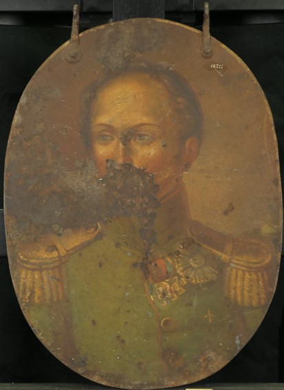
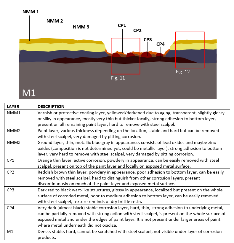
The iron support is in satisfactory condition and is stable and solid. In places where the paint layer has fallen off, there is a fairly thick layer of passivated corrosion present, except on small local areas of fresh damage, where the paint peels to the ground layer. Corrosion is mostly inactive and stable, there are local smaller active corrosion areas. Corrosion products from the support locally penetrate the paint layer where they cause discoloration. If the paint layer is removed, thin layer of dark brown (almost black) corrosion products can be seen underneath it. Locally, underlaying thicker deposits of corrosion products are causing paint layer to swell.
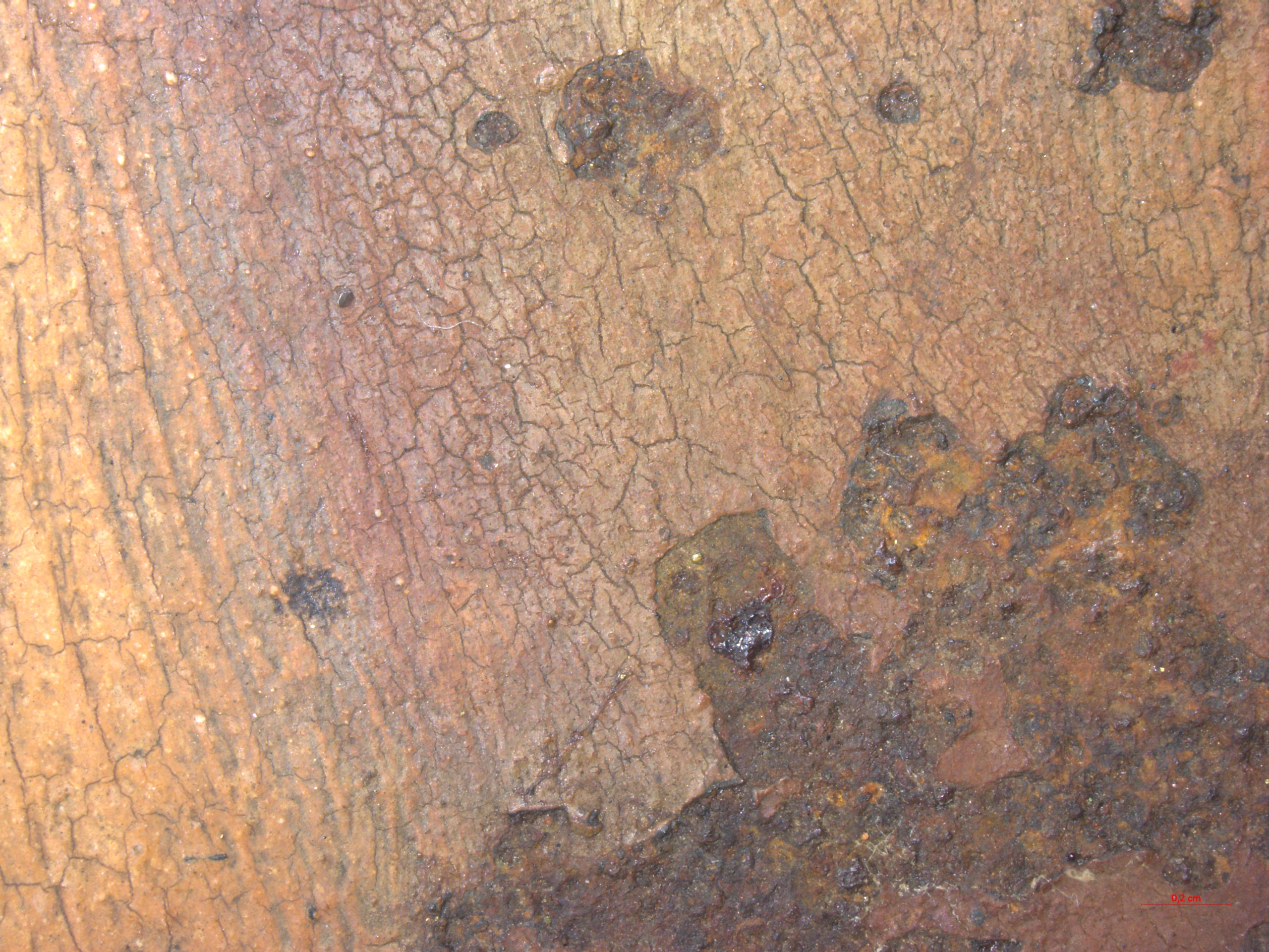
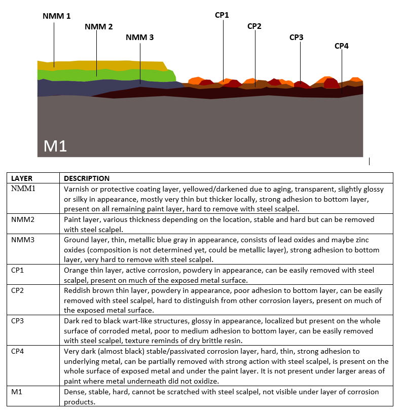
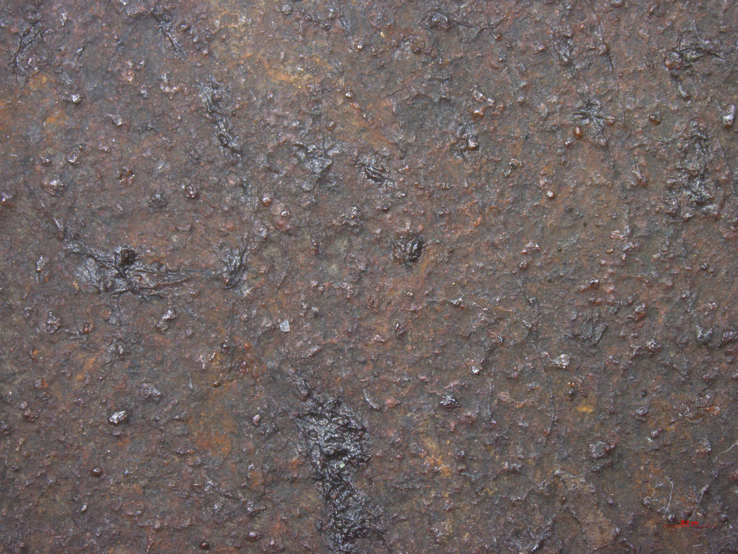
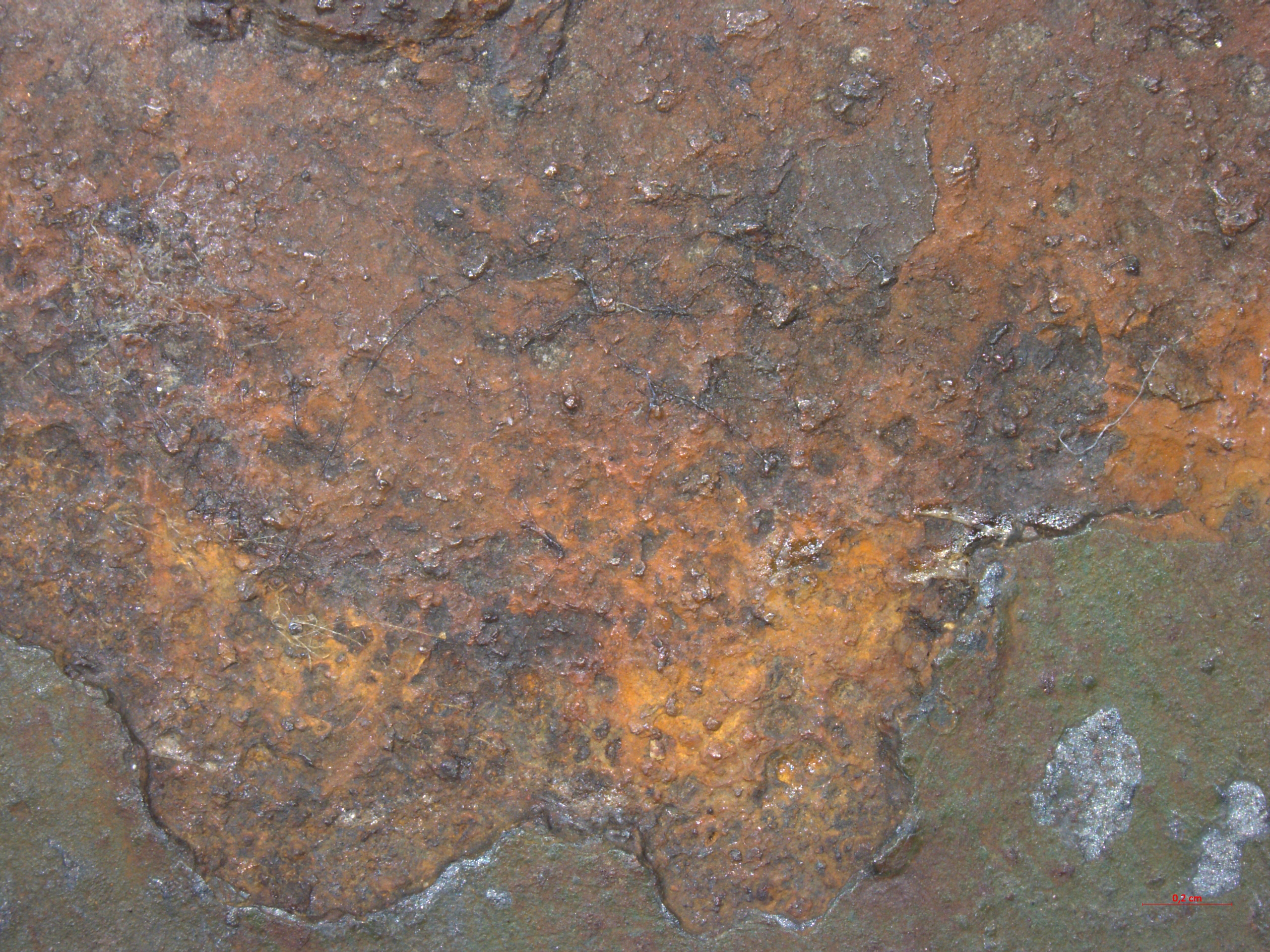
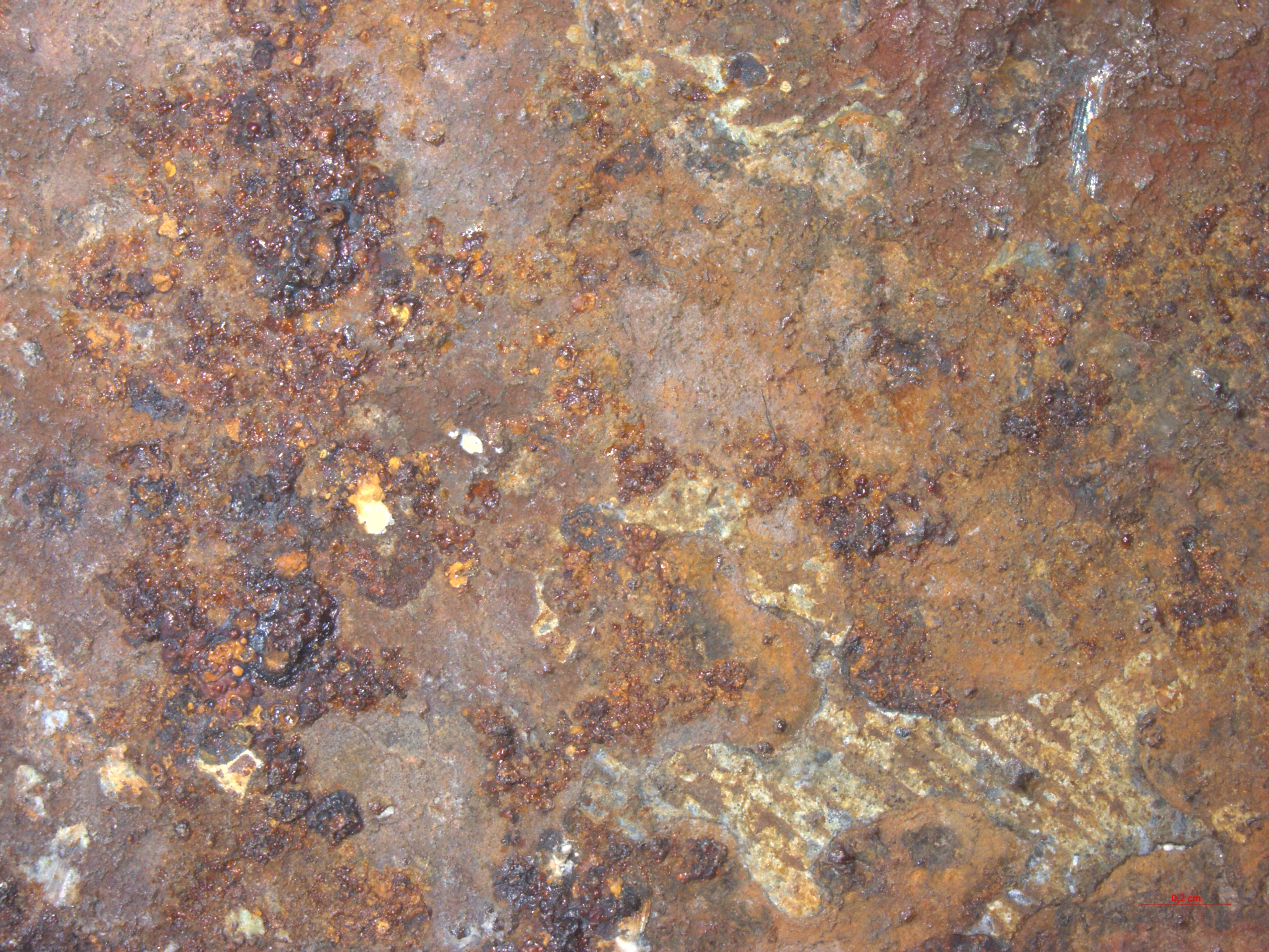
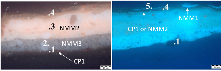
On the front side of the painting, corrosion products are heterogenous and form multiple layers. Statigraphy of corrosion products is similar on most areas and can be divided by four main layers: outer layer of localized brittle, powdery orange products (CP1), followed by reddish brown powdery layer (CP2), followed by dark red (almost black) shiny brittle wart-like structures of different sizes that appear indvidually but cover most of the surface (CP3). The lowest layer consists of dark brown, almost black thin hard layer that covers original metal surface (CP4).(Fig. 5) Paint surface on the bottom part of the painting was cleaned with cleaning systems that contained water (rigid agarose gels with chelators) that locally came into contact with exposed corroded metal; on those areas bright orange active corrosion (flash corrosion) formed almost immidiately after removal of rigid gels (CP1). (Fig.6).
On the back side, corrosion on the upper area of the painting are the same type as on the front side. (Fig. 7) On the lower area, corrosion type is slightly different: extensive pitting corrosion appears that forms deep individual pits across the whole surface of that area. Corrosion statigraphy is similar as on the front side of the painting: orange, brittle corrosion products (CP1) are located mainly around the pits and inside them on top of other corrosion layers, but also on top of the remaining paint layer - this is probably because of cleaning of the remaining paint layer with agarose gels which contain water (same problem as on the front side of painting). Inside the pits underneath CP1 is a layer of brownish-red brittle corrosion products (CP2), which is hard to distinguish from other layers. Next layer are dark red (almost black) shiny brittle wart-like structures (CP3) and underneath a thin, hard layer of very dark brown products (CP4). (Fig. 8).
Based on the type and colour of corrosion products we can assume that they are mostly iron oxides, with iron hydroxides appearing locally where the surface was in contact with water. This description is based solely on visual analysis. We can assume that the corrosion is atmospheric since the layer is thin and stable and the object was originally kept outside. A sample of corrosion products was taken for XRD analysis in hopes of more precise identification or confirmation of these assumptions (currently awaiting results).
None
None
None.
None.
The painting is made from iron alloy plate. Precise microstructure of the metal has not been analysed since metallographic analysis was not executed. Based on the pigments used for the paint layer the object dates to late 19th century. Areas with missing paint layer appear heavily corroded but the layer of atmospheric corrosion is thin and the metal underneath is in good and stable condition. Various layers of corrosion can be observed visually, mostly iron oxides and hydroxides. Corrosion products are also present on top of the paint layer.
References on object and sample
References object
1. Milič, Z., 'Konserviranje in restavriranje železa 3.1.1', Skupnost muzejev Slovenije, 2001, p. 1-8, http://www.sms-muzeji.si/ckfinder/userfiles/files/udatoteke/publikacija/netpdf/3-1-1.pdf (accessed 1 September 2023).
2. H. Pucelj Kranjc, Konserviranje-restavriranje oljne slike na železni pločevini Požar v Kranju iz Narodnega muzeja Slovenije, MA diss., University of Ljubljana, 2018, https://repozitorij.uni-lj.si/IzpisGradiva.php?id=105959&lang=slv (accessed 1 September 2023).
3. D.A. Scott and G. Eggert, Iron and steel in art, London, Archetype Publications, 2009, p. 107-122.
References on analytic methods and interpretation
4. D.A. Scott and G. Eggert, Iron and steel in art, London, Archetype Publications, 2009, p. 35-52.