Figurine of Egyptian God Bes 1 - ÆIN 223
Ida. Langemark (The Royal Danish Academy, Copenhagen, Capital Region, Denmark)
 Credit The Royal Danish Academy, I.Langemark.
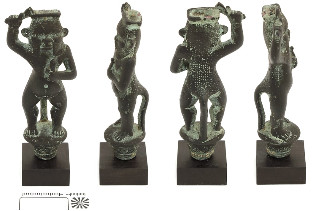
Bes appears in his typical form with grotesque face, naked male body, short and hooked legs, lion's mane, mouth, and tail. The god was perceived as a protector who defeated evil forces with force of arms or magical music and is here depicted as a snake slayer with a sword raised in his right hand and a cobra in his left hand. Dimensions: Height=11.6cm; Width=4.8cm; Thickness=3.3cm.
The figure has lost its crown, which was shaped like a feather tassel, but its original existence is revealed by an empty mounting hole at the top of the head. The figure originally stood on vertical stands that illustrated the flowering papyrus stalks, symbols of health and freshness, wadj, but only sparse remains are preserved under the figure's feet. The wadj-symbol with the god on top may have been mounted at the top of a ceremonial staff or had a place on a base, possibly together with other figures (Jørgensen 2009 23.1.).
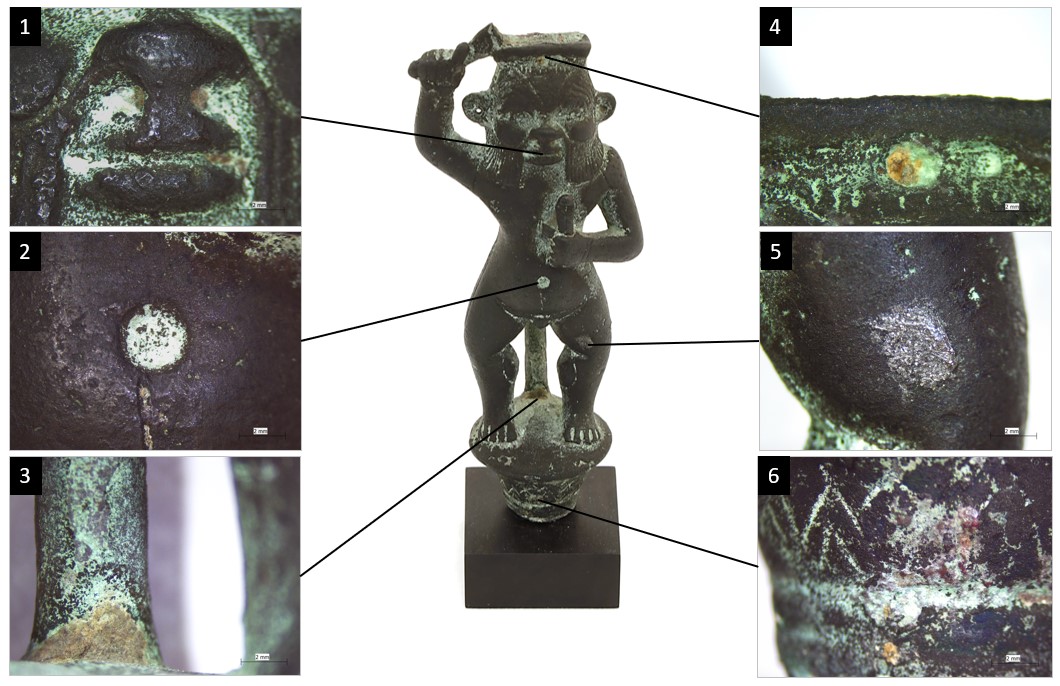
The figure is covered by a dark corrosion layer which could be an original artificial patina, with light green powdery corrosion products located in and around engravings, edges, and curves, as well as horizontal surfaces and the insides of legs and tale (Fig. 2 and 3).
Votive figure
Archaeological, ancient Egypt, exact provenance unknown
Recovered late 19th century
3rd Intermediate Period to Late Period
None.
Unknown
Ny Carlsberg Glyptotek (Hast Rebecca), Copenhagen
Ny Carlsberg Glyptotek (Hast Rebecca), Copenhagen
ÆIN 223
N/A
Mounted to wooden plinth.
Inventory number applied in gold ink of unknown composition.
 Credit The Royal Danish Academy, I.Langemark.
 Credit The Royal Danish Academy, I.Langemark.
 Credit The Royal Danish Academy, I.Langemark.
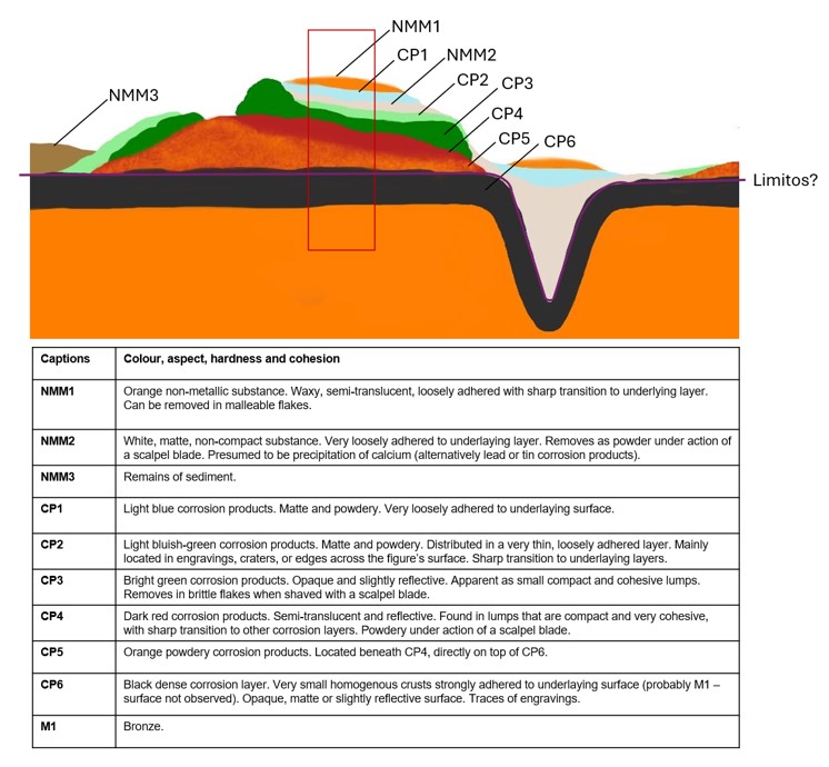
Stratigraphic representation:
The schematic representation below gives an overview of the corrosion structures encountered on the figurine from a first visual macroscopic observation.
Seven samples of corrosion products and non-metallic material were taken and observed under polarized microscopy (Fig. 9), two samples (NMM2) were examined with SEM-EDS and 1 sample under P-XRD.
Leaded Bronze
Cast
The Royal Danish Academy (Langemark Ida), Copenhagen, Capital Region
The Royal Danish Academy (Langemark Ida), Copenhagen
March 8th 2024, chemical and molecular analyses of corrosion products
None.
Analyses performed:
Non-invasive approach
- Portable micro-X-ray fluorescence (μ-XRF) on different measuring locations across the bronze surface (Fig. 7). Measurements were made with a portable Bruker Tracer 5G XRF, with an analysis time of 30 sec and a spot size of 8 mm, mode general metal.
Invasive approach
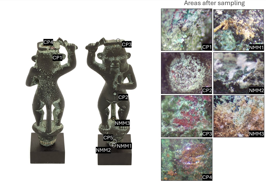
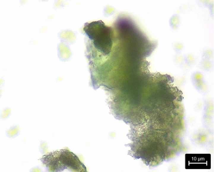
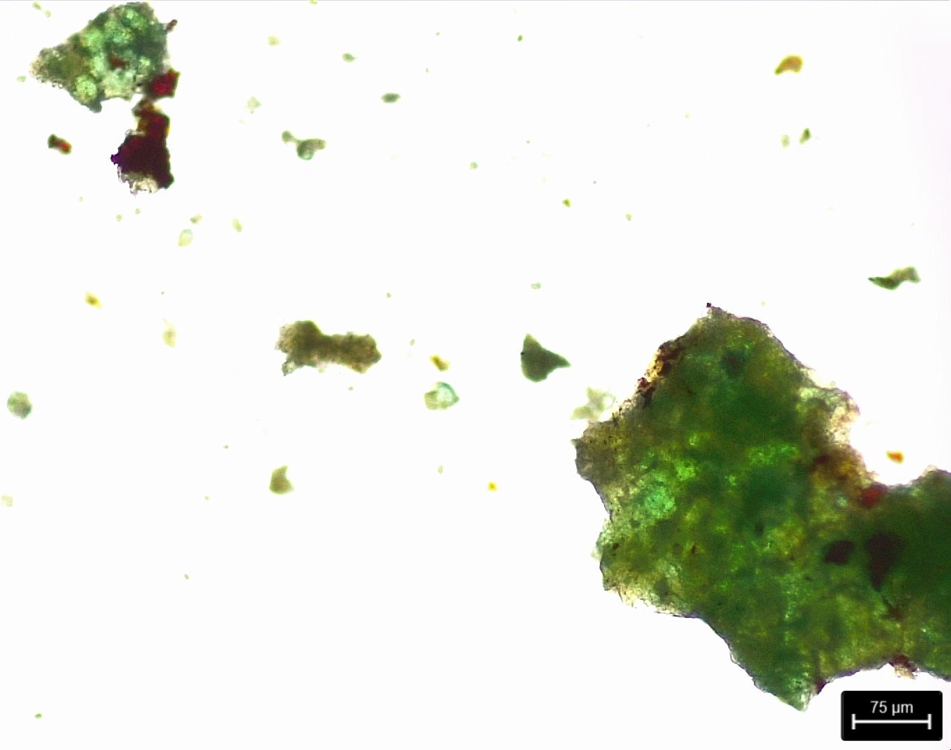
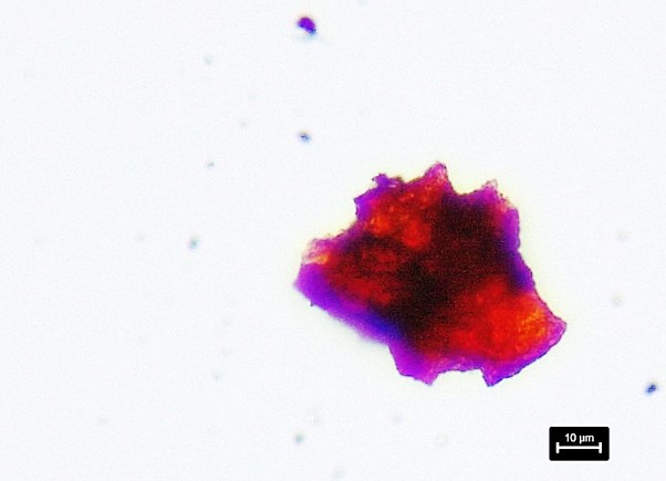
- Polarized microscopy on 4 corrosion samples (CP1-CP4) and 3 samples from non-metallic compounds (NMM1-NMM3)(Fig. 10).
A small amount (1>mg) of various corrosion types was removed from the surface with scalpel under binocular microscope. The corrosion powder was placed in a drop of Euparal, covered with an object glass, and left to dry for some days. The corrosion samples were observed with a Leica 750P-microscope, and micrographs obtained with the software LAS X.
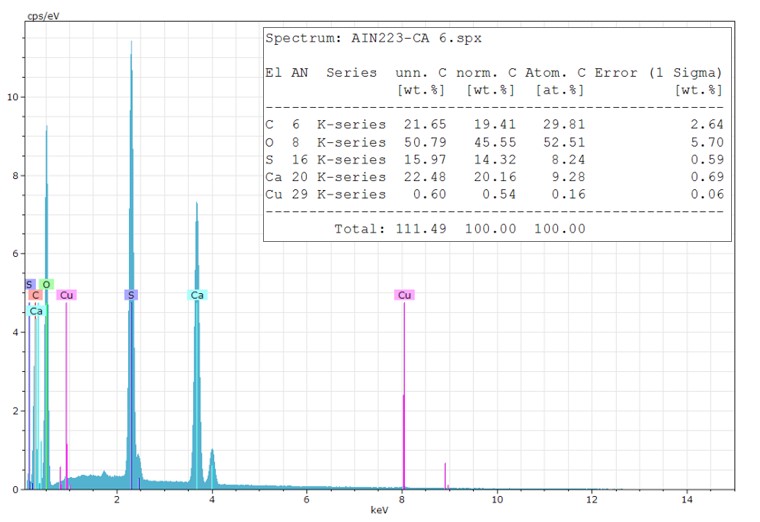
- Scanning electron microscopy /Energy Dispersive X-ray Spectrometry (SEM-EDS) on corrosion powder samples of NMM1 and NMM2. Samples was prepared by scraping of material from the surface with scalpel under binocular microscope. The sample material was attached to steel pins with double sided carbon tape. Analyses were performed with a Hitachi S - 3400N SEM-EDS, 15,0 kV and 300 sec.
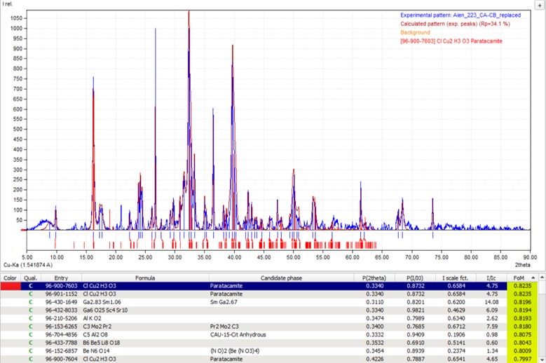
- Powder X-Ray diffraction (PXRD) on corrosion products sampled (CP1 and CP2). Due to the amount of material required for PXRD-analysis, sampling was done on all areas with a sufficient amount of corrosion products. PXRD-analyses were performed with a PANalytical X’Pert Pro diffractometer at the Faculty of Farmacy, Physics and Chemistry, University of Southern Denmark. Measurements were obtained with the software Data collector, and subsequent data processing was done with the applications HighScorePlus and Match! respectively.
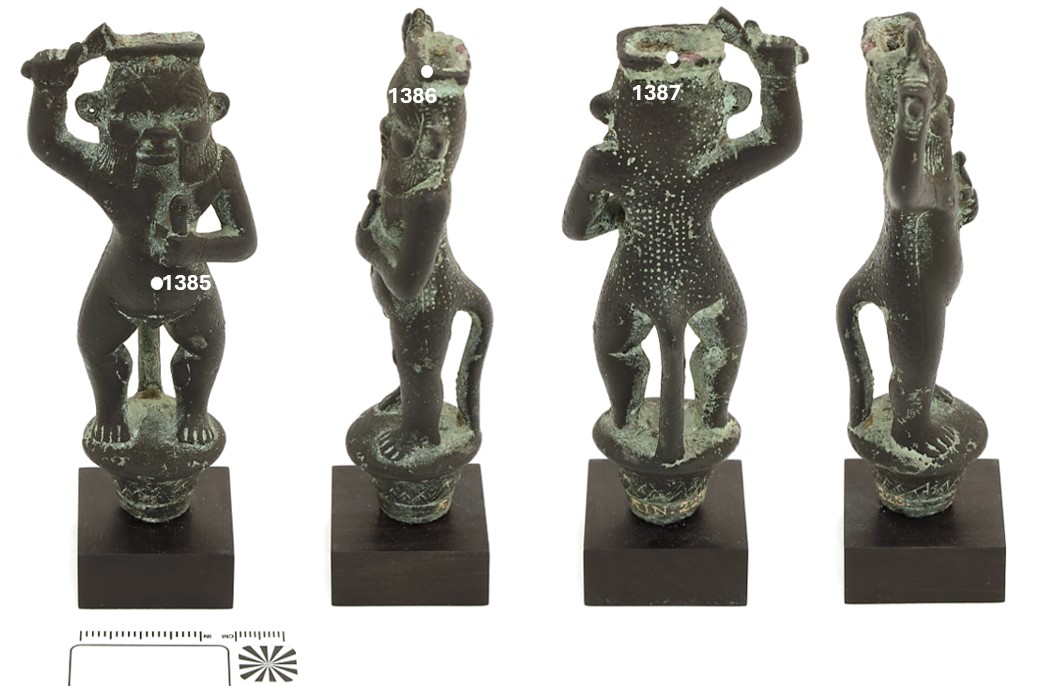
The μ-XRF-measurements have been performed on un-polished surface areas. They should be seen as indicative. Still the metal is a leaded bronze.
| Measurement no. | Cu | Pb | Sn | Sb | Ag | Zn | Ni |
| 1385 | 71.3 | 19.6 | 9.2 | < | < | < |
< |
| 1386 | 81.8 | 16.9 | 1.3 | < | < | < | < |
| 1387 | 72.4 | 20.3 | 7.3 | < | < | < | < |
Table 1: Chemical composition (Elements mass (%)) of the figurine in the areas located on Fig. 7.
The μ-XRF-analysis shows a content of approx. 19 % (in weight) lead as an average of measurement 1385, 1386, and 1387. The lowest tin-content of 1.3 wt% Sn is seen in measurement 1386 (crown, red corrosion products), the highest of 9.2 wt% Sn in measurement 1385 (powdery corrosion products accumulated in the navel).
Based on the figure’s visual dark appearance, traces of elements commonly associated with artificial patination of archaeological bronze, such as gold (Au) and silver (Ag), was expected to be found in the metal alloy (Benzonelli 2017, Berger 2015, Mohamed & Darweesh 2012). However, the performed measurements showed no trace of gold, silver, or other alloying elements.
None.
None
Cu
Sn, Pb
None.
The heterogenous corrosion crust covers the whole object with various types of corrosion products, mostly Robbiola type I. Most of the bronze surface is covered with a dark grey patina, with powdery light green corrosion products located in and around engravings, edges, and curves, as well as horizontal surfaces and the insides of both legs and tail.
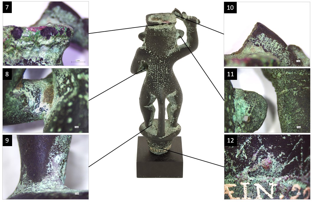
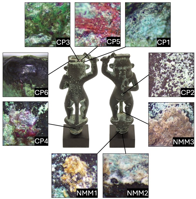
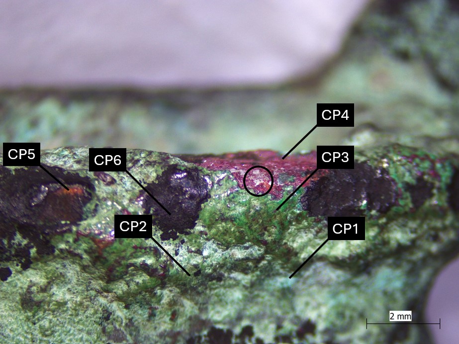
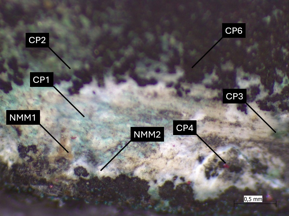
Six corrosion products and three non-metallic materials have been identified and can be visualised on Fig. 4 and as stratigraphies on the crown (Fig. 5) and the standing base (Fig. 6) respectively. As a reminder:
- CP1: light blue corrosion product found on a few localized spots,
- CP2: a thin corrosion layer consisting of light green powdery corrosion products (Fig. 11),
- CP3: a dense bright green layer found at a few localized spots (Fig. 12),
- CP4: a hard, brittle red layer (Fig. 13),
- CP5: powdery orange corrosion products located beneath CP4.
- CP6: a dense black/dark grey, very hard corrosion layer, that contains corresponding markers to the limit of the original surface. In some areas the sequence has an unevenness surface (with a mattifying effect), in other areas the surface is more regular and more reflective. At some locations on the crown, the original surface has been replaced by structural voids in form of small craters.
PXRD-analysis of corrosion powder obtained from CP1 and CP2 show the presence of the copper tri-hydroxychloride paratacamite (Fig. 14), and traces of spertiniite (full data-sheet available on request).
SEM-EDS-analysis of the white substance found on the standing base (NMM2) shows a high content of calcium (Fig. 15). In some areas, the white layers have an almost waxy appearance, a feature that often reveals the presence of nantokite (copper oxychloride). Distinguishing between areas of calcium precipitation and nantokite would require more samples to be analysed.
 Credit The Royal Danish Academy, I.Langemark.
 Credit The Royal Danish Academy, I.Langemark.
 Credit The Royal Danish Academy, I.Langemark.
 Credit The Royal Danish Academy, I.Langemark.
Multiform
Mostly type I with locally type II (Robbiola)
Small amounts of white and waxy corrosion product have been observed (encircled in Fig. 5). Based on the presence of high amounts of copper tri-hydroxychloride (paratacamite) in the sample obtained from CP1 and CP2, this substance could very well be nantokite (CuCl).
None.
The figurine is a leaded bronze.
The corrosion products, which cover the entire object, are multi-layered. Most of the corrosion layers correspond to type I of Robbiola.
The presence of copper tri-hydroxychloride paratacamite was detected by PXRD in a sample of corrosion powder obtained from CP1 and CP2. PXRD analysis also detected small amounts of spertiniite, which is consistent with the visual aspects of CP1. However, it could also be the formation of chalconatronite (Na2Cu(CO3)2 - 3H2O), a corrosion product associated with archaeological bronze from Egypt (Frondel 1955). CP3 is thought to consist mainly of malachite, CP4 and CP5 of cuprite. The dark corrosion layer CP6 might consist of tenorite as an original artificial patina.
Three non-metallic compounds were found on the figurine: wax remains (NMM1), calcium precipitation confirmed by SEM-EDS (NMM2) and sediment remains (NMM3).
References on object and analytical methods
Reference object
1. Jørgensen, M. (2009) Katalog Ægypten V. Ny Carlsberg Glyptotek 2009, 23.1.
Reference analytical method
2. Benzonelli, A., Freestone, I. C., Martinón-Torres, M. (2017) A better shade of black: effects of manufacturing parameters on the development of ancient black bronzes in Archaeometry, Volume 59, Issue 6, 1034-1049. DOI: 10.1111/arcm.12299.
3. Berger, D. (2015) Artificial patination in Early Iron Age Europe: an analytical case study of a unique bronze artefact, in Journal of Archaeological Science, 57, 2015, 130-141. DOI: 10.1016/j.jas.2015.01.025.
4. Frondel, C., Gettens, R.J. (1955) Chalconatronite, a new mineral from Egypt in Science, 122, 75-76.
5. Mohamed, W. & Darweesh, S. (2012) Ancient Egyptian Black-Patinated Copper alloys, in Archaeometry, 54, 175-192.