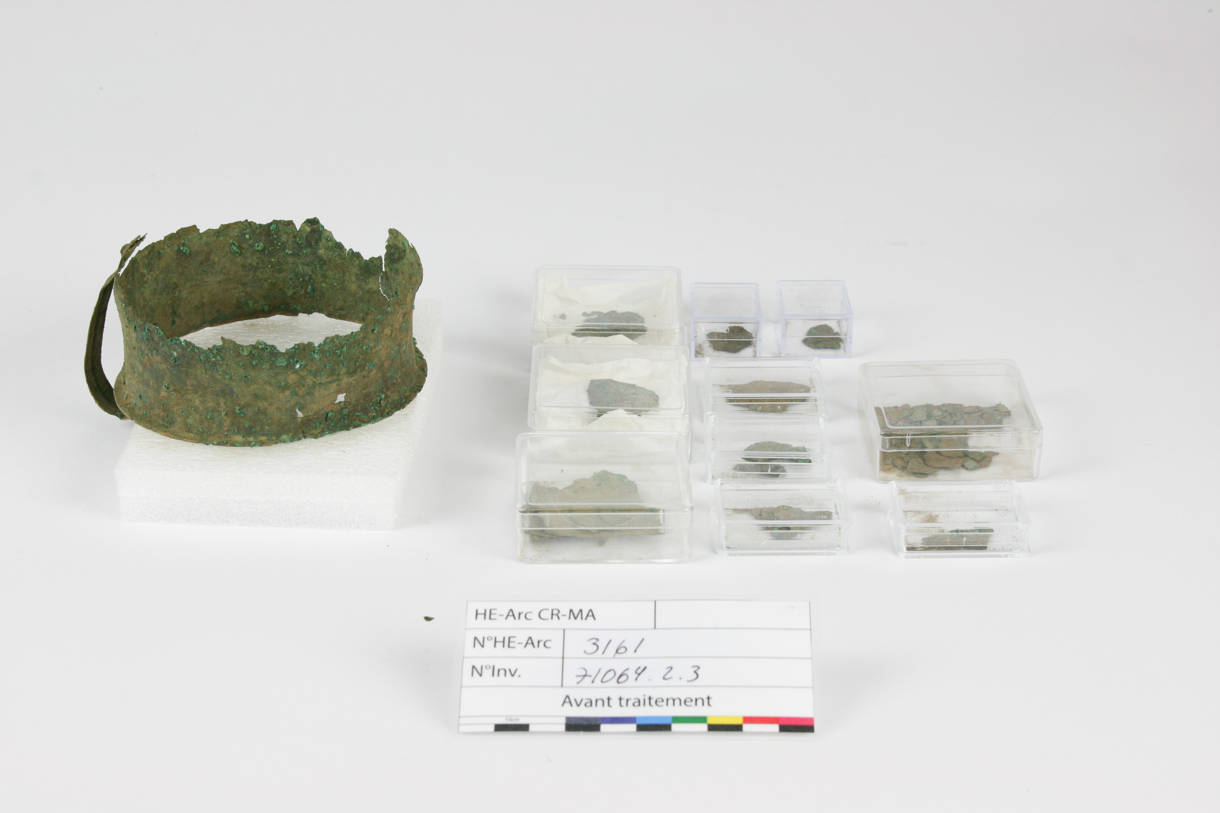
Jar/cup FO-Nr. 71064.2.3
Ingrid González. (HE-Arc CR, Neuchâtel, Neuchâtel, Switzerland) & Valentin. Boissonnas (HE-Arc CR, Neuchâtel, Neuchâtel, Switzerland)
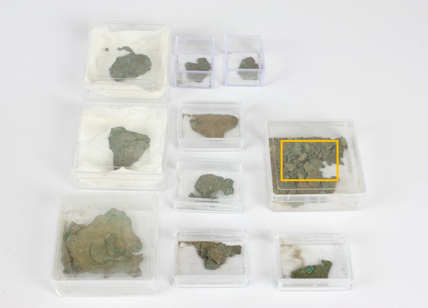
Jar/cup (Figs. 1 & 2) developing locally green corrosion products covered with sediments and some organic remains presenting a filiform structure. The object preserves its central cylindric main body. The bottom has been completely detached but several fragments are preserved (Fig. 3). Dimensions: H = 7cm; D = 12cm ; T = 0,3mm. The object comes from a cremation tomb.
Cup
Castaneda, Grisons, Switzerland
Excavation 2021, grave 1
Late Iron Age
None.
Soil
Archaeological Service of the Canton of Grisons, Grisons
Archaeological Service of the Canton of Grisons, Grisons
FO-Nr. 71064.2.3
N/A
None.
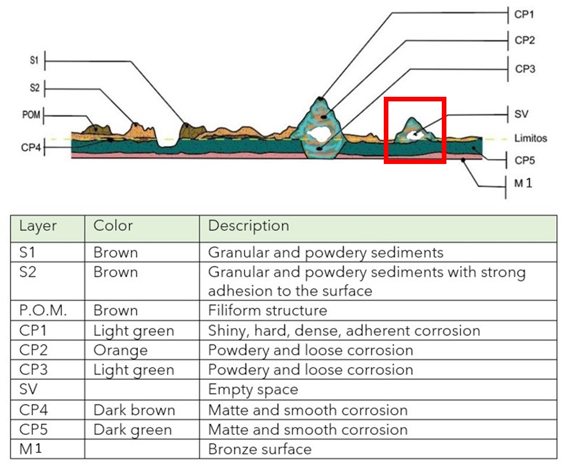
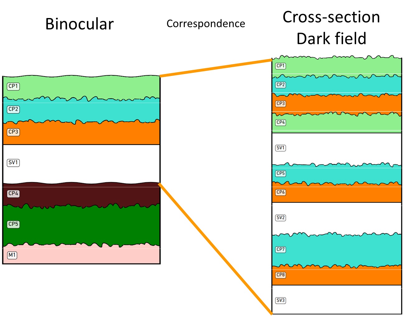
The schematic representation below gives an overview of the corrosion structure encountered on the jar/cup from a first visual macroscopic observation.
 Credit HEI Arc, C.Cséfalvay.
The cross-section corresponds to a fragment detached from the base of the cup/jar (Fig. 3). Its corrosion structure is representative of the corrosion products and the metallic core observed on the whole cup/jar.
Bronze
Hammered, turned and final cold work
Tasse_Mag
Archaeological Service of the Canton of Grisons, Grisons
Archaeological Service of the Canton of Grisons, Grisons
2023, metallography and chemical analyses
None.
Analyses performed:
Non-invasive approach
- XRF with handheld portable X-ray fluorescence spectrometer (NITON XL3t 950 Air GOLDD+, Thermo Fischer®). General Metal mode, acquisition time 60s (filters: Li20/Lo20/M20).
Invasive approach (on the sample)
- Optical microscopy: the sample is embedded in an EPOFIX resin and polished, observed then with a digital microscope KEYENCE VHX-7000 in bright and dark field.
- Metallography: the polished sample is etched with alcoholic ferric chloride and observed by optical microscopy in bright field.
- SEM-EDS: the sample is coated with a carbon layer and analyses are performed on a SEM-FEG JEOL 7001-F equipped with a silicon-drift EDS Oxford detector (Aztec analysis software). Accelerating voltage is of 20 kV and probe current is at about 9 nA. The relative error is considered of about 10% for content range <1mass%, and of 2% for content range of >1mass%.
- FTIR: the sample of a cluster was characterised on a Perkin Elmer System 2000. Spectra were measured in transmission mode at a spectral resolution of 4 cm-1. The sample was flattened on a diamond cell for the analysis.
The XRF analysis of the surface of the cup/jar was carried out on the main body and the handle (Fig. 2). The metal is presumably a copper-tin alloy with some lead, while the light elements detected are from the sediments (Si and Al). The presence of phosphorus (P) could come from human remains from the tomb. There is a very slight presence of titanium (Ti) which could be a kind of contamination.
| Elements (wt%) | Cu | Sn | Pb | Ti | Al | P | Si |
| Main body | 41 | 41 | 0.5 | 0.5 | 7 | 6 | 3 |
| Handle | 45 | 24 | <0.5 | <0.5 | 15 | 3 | 12 |
Table 1: Results of the XRF analysis of the surface of the cup. Method of analysis: handheld portable X-ray fluorescence spectrometer, HE-Arc CR.
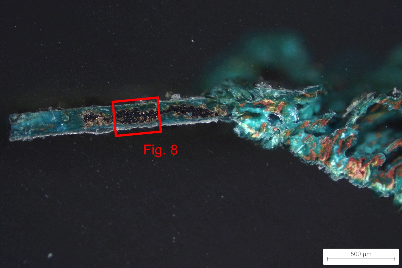
The EDX analysis (Table 2) of the residual metal on cross-section indicates that it is a tin bronze (12 wt% Sn) with a low percentage of lead (0.1 wt% Pb). Clearly the XRF analysis shows a surface enrichment with tin.
The etched metal shows a structure of polygonal grains with strain lines (Fig. 8). This indicates that either the object was submitted to a light annealing process after the intense mechanical work that can be perceived through the slip lines. Or the final mechanical work did not affect the main structure of the grains formed by the previous annealing. Small Pb inclusions are distributed through the sample.
| Elements (wt%) | Cu | Sn | Pb |
| Core metal | 88 | 12 | 0,1 |
Table 2: Chemical composition of the core metal. Method of analysis: SEM-EDS.
Polygonal grains and strain lines
Cu
Sn, Pb
None.
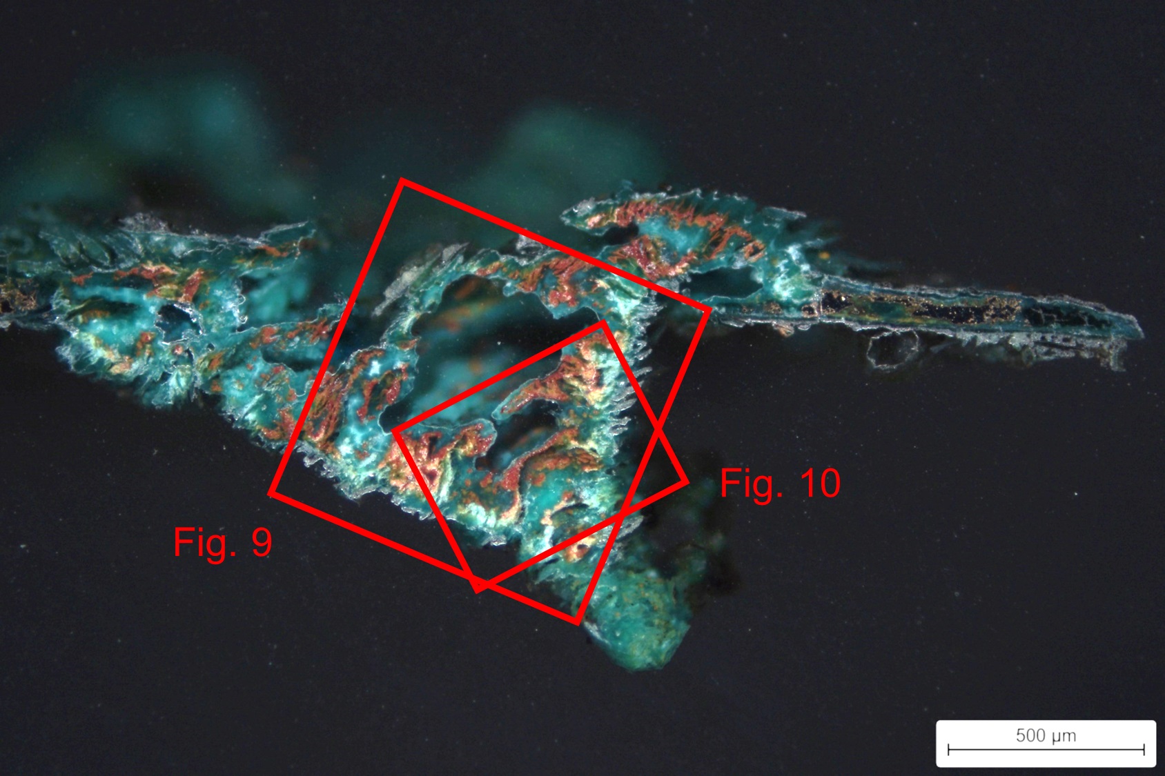
The observation of a pustule in cross-section in dark field shows three main corrosion products overlaying each other (Fig. 9). The first green corrosion layer (CP1) possibly corresponds to atacamite (Cu2Cl(OH)3) as suggested by the FTIR spectrum of Fig. 10. The second corrosion layer inside the pustule (CP2) is probably a copper oxide - cuprite (Cu2O) and the third light green (CP3) corrosion layer could be malachite (Cu2CO3(OH)2), as suggested again by FTIR spectrum of Fig. 12.
The multilayer pustule corrosion has developed similar to the process presented by Formigli (1975) and Scott (2002).
Elemental EDX analysis and mapping (Fig. 10) show phases which are richer in Sn than in Cu. Cl is located in the area closest to the outside of the pustule. Phosphorus (P) is also located in this area, possibly as a soil contaminant in the presence of human remains.
 Credit HE-Arc CR.
 Credit SIK-ISEA, A. Vichi.
 Credit SIK-ISEA, A. Vichi.
Multiform (warty - uniform) - pitting
Both Formigli (pustules) and type I (Robbiola) otherwise
None.
The stratigraphies obtained by binocular and cross-section observation show a few differences that can be attributed to the different scales of observation and the impossibility to visualise both a pustule and the remaining metal underneath. Under binocular microscope, it is difficult to differentiate the multiple strata of the pustule. Only the main strata are indicated.
Under cross-section, the distribution of strata appears more clearly while this distribution and succession of strata are different in the middle of the pustule versus its edges. CP4 is similar to CP1, while CP5/CP7 are similar to CP2 and CP6/CP8 are similar to CP3.
CP4 and CP5 of the Bi stratigraphy do not show up on the CS stratigraphy. Similarly the M1 stratum is not described.
The jar/cup is made of a tin bronze with Pb inclusions. Metallographic observation of the remaining metal revealed a polygonal grain structure with strain lines. This indicates that the metal was annealed and cold worked several times, with a final cold working. The metal is in an advanced state of mineralisation with the formation of pustules crossing the full thickness of the metal.
Observation of the corrosion layers showed a typical corrosion structure for an archaeological bronze. The main corrosion products found are malachite and most likely cuprite and atacamite. The object shows a Sn enrichment process on the surface. The presence of P can be attributed to the presence of human remains in the grave, while the presence of Si, Al and Ti is mainly due to the elements of the burial soil.
The limit of the original surface limit corresponds to the upper surface of the brown and dark green layer. The corrosion is multiform type I according to Rabbiola (1998) while the pustules are of the Formigli type (Formigli 1975).
References on object and sample
1. González, I. (2023) Tasse FO-Nr. 71064.2.3, Archaeological Service of the Canton of Grisons, rapport d’intervention. Haute Ecole ARC, Neuchâtel, non-publié.
2. MiCorr_Fragments of oenochoe GV132-01/US26-OBJ.10.
3. González, I. (2023) Jar/cup - Bronze - Late Iron Age, FO-Nr. 71064.2.3, MiCorr, Switzerland, Haute Ecole ARC, Neuchâtel, non-publié.
References on analytic methods and interpretation
4. Formigli, E. (1975) « Die Bildung von Schichtpocken auf antiken Bronzen ». In ArbeitsblätterAdR, Gruppe 2, 51-58.
5. Robbiola, L. (1998) Morphology and mechanisms of formation of natural patinas on archaeological Cu-Sn alloys. In Corrosion Science, 40, 12, 2083-2111.
6. Scott, D. A. (2002) Copper and bronze in Art, corrosion, colorants, conservation. Getty publications, Los Angeles.
7. Scott, D. A. (1991) Metallography and microstructure of ancient and historic metals. Getty publications, Los Angeles.