Blade fragment of a winged axe FK43
Marianne. Senn (EMPA, Dübendorf, Zurich, Switzerland) & Christian. Degrigny (HE-Arc CR, Neuchâtel, Neuchâtel, Switzerland)
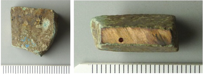
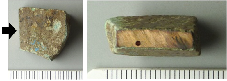
Blade fragment of a semi-finished median-winged axe. Its surface is covered with a thick dark green corrosion crust (Fig. 1). Dimensions: L = 20mm; Tmax. = 8.5mm; WT = 15g.
Tool
Obstgartenstrasse, Erlenbach, Zurich, Switzerland
Excavation 1980.002
Middle Bronze Age
1550BC _ 1350BC
Soil
Kantonsarchäologie, Dübendorf, Zurich
Kantonsarchäologie, Dübendorf, Zurich
FK43
Not conserved
Nothing to report.
Stratigraphic representation: none.
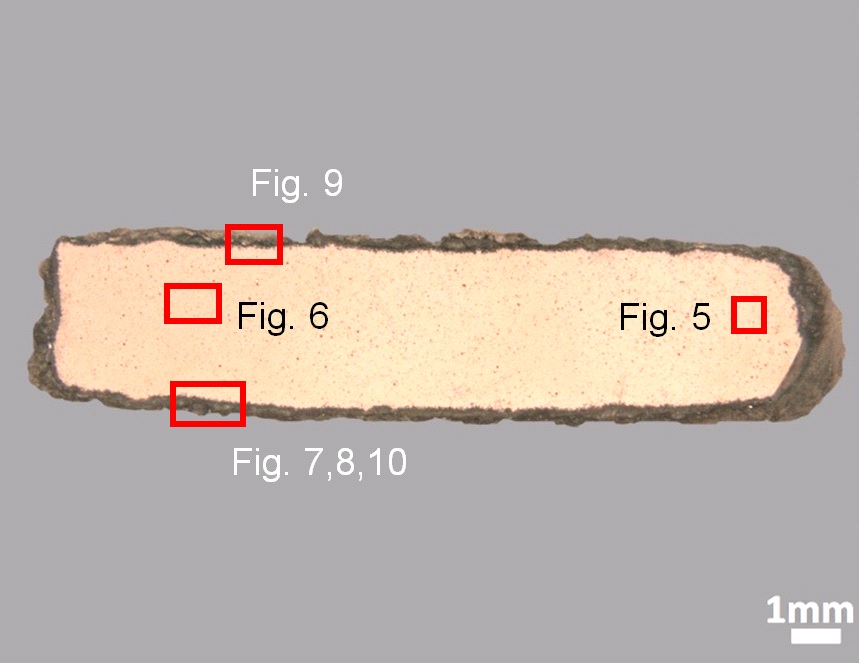
The sample was cut from the fragment shown in Fig. 2. The cross-section is rectangular in shape (L = 17mm, W= 4mm) and has a thick corrosion crust (Figs. 2 and 3).
Tin Bronze
As-cast
ERL-43
Begbroke Science Park (Peter Northover), Yarnton, England
Kantonsarchäologie, Dübendorf, Zurich
Date unknown, metallography and chemical analyses
Nothing to report.
Analyses performed:
Metallography (etched with ferric chloride reagent), Vickers hardness testing, SEM/EDS, EPMA/WDS, Raman spectroscopy.
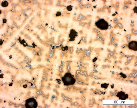
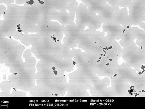
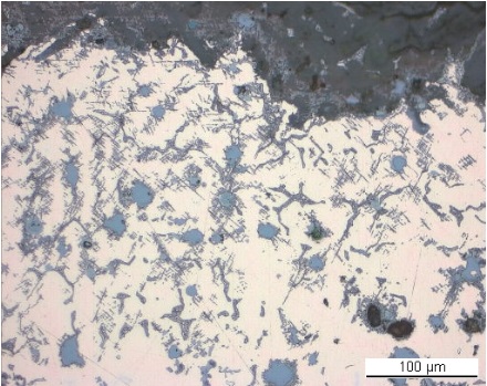
The remaining metal is a tin bronze (Table 1) with high porosity and grey copper sulphide inclusions (Figs. 5 and 6, Table 2). The etched metal has the typical dendritic structure of a cast tin bronze with an average hardness of HV1 135 (Fig. 6). The cored dendritic structure is surrounded by an alpha-delta eutectoid. The core of the dendrites is rich in Cu whereas the outer layers are rich in Sn.
| Elements | Cu | Sn | As | Fe | Ni | Pb | Sb | Co | Ag | Au | Zn | Bi | Si |
|---|---|---|---|---|---|---|---|---|---|---|---|---|---|
| mass% | 85.14 | 11.95 | 1.54 | 0.49 | 0.39 | 0.18 | 0.14 | 0.13 | 0.02 | 0.02 | < | < | n. d. |
Table 1: Chemical composition of the metal. Method of analysis: EPMA/WDS, Lab Department of Materials, University of Oxford.
|
Elements |
Cu | S | Fe | Total |
|---|---|---|---|---|
| Dark-grey inclusion | 66 | 24 | 10 | 100 |
Table 2: Chemical composition (mass %) of the dark-grey inclusions seen in Fig. 5. Method of analysis: SEM/EDS, Laboratory of Analytical Chemistry, Empa.
 Credit HE-Arc CR.
Dendritic structure + strain lines (metal surface)
Cu
As, Sn
Nothing to report.
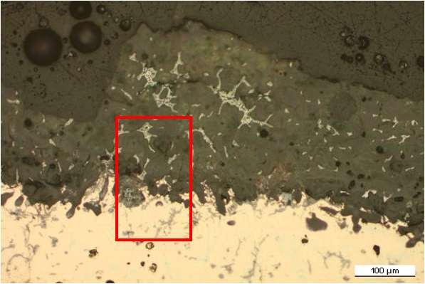
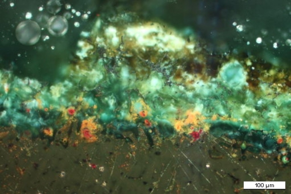
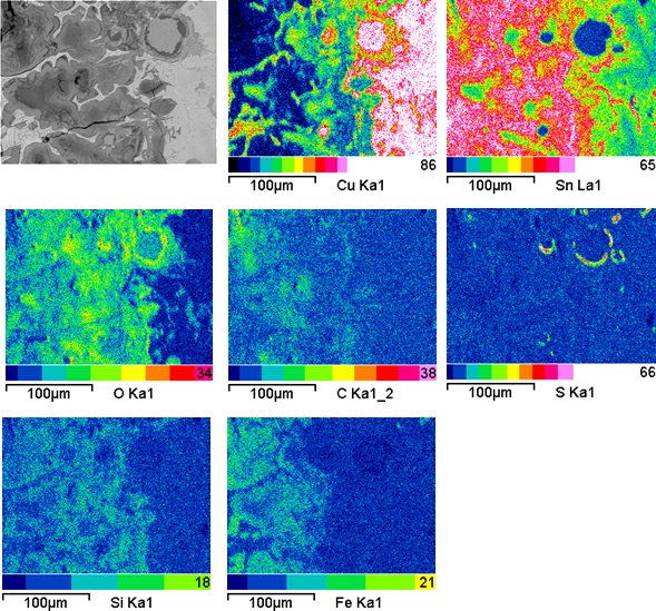
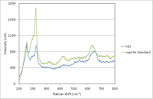
A dark green corrosion crust with a thickness between 100 and 320µm covers the entire surface of the blade fragment (Fig. 7). It retains a metallic ghost structure (Sn-rich eutectoid alpha + delta phase). Under polarized light localized orange and red corrosion products can be seen at the metal - corrosion crust interface (Fig. 8). Interdendritic corrosion and corroded slip lines can be seen in the metal structure and near fissures (Fig. 9). Elemental mapping (Fig. 10) shows that the green layer is Sn-rich (CP1, probably cassiterite, SnO2) and depleted of Cu, whereas the orange and red corrosion particles are depleted of Sn and rich in Cu (Fig. 10, Table 3). Their Raman spectra indicate that they are mainly composed of cuprite (Fig. 11). The overall corrosion crust contains O, Si, C and Fe from the environment, while S is concentrated around the cuprite particles (Fig. 10).
|
Elements |
O | Si | P | Fe | Ni | Cu | As | Sn | Total |
|---|---|---|---|---|---|---|---|---|---|
| Surface CP1 | 43 | 0.8 | < | 6.2 | < | 16 | 1 | 43 | 111 |
| Middle CP1 | 42 | 1.7 | 0.7 | 12 | < | 10 | 0.7 | 43 | 110 |
| Red/orange CP in CP1 | 41 | 0.9 | < | 4.4 | < | 36 | < | 22 | 104 |
| Remnant metal phase | 9 | 0.7 | < | 5 | 0.8 | 34 | < | 47 | 97 |
Table 3: Chemical composition (mass %) of the corrosion crust from Fig. 7. Method of analysis: SEM/EDS, Laboratory of Analytical Chemistry, Empa.
 Credit HE-Arc CR.
 Credit HE-Arc CR.
 Credit HE-Arc CR.
 Credit Empa.
 Credit SNM.
Fig. 11: Raman spectrum of a red-orange corrosion particle (S42) of Fig. 8 compared to the cuprite standard spectrum. Settings: laser wavelength 532nm, acquisition time 10s, one accumulation, filter D2 (0.75-0.8mW), hole 500, slit 80, grating 600. Method of analysis: Raman spectroscopy, Lab of Swiss National Museum, Affoltern a. Albis ZH,
Uniform - selective
Type I (Robbiola)
Nothing to report.
Corrected stratigraphic representation: none.
The evenly corroded tin bronze contains numerous sulphide inclusions and shows signs of interdendritic corrosion penetrating the metal structure. The Sn enriched surface is decuprified and polluted by the environmental elements such as O, Si, Fe, C, Al and Cl. The corrosion crust is composed mainly of a dark green layer with local orange-red cuprite particles at the interface with the remaining metal. Both the remnant metallic phases and the Sn-rich corrosion layer can be interpreted as inferior markers, defining the limit of the original surface which is located above. For the above mentioned reasons, the corrosion is thought to be of type 1 according to Robbiola et al. 1998.
|
References on object and sample |
|
Reference object 1. Fischer, C. (1997) Innovation und Tradition in der Mittel- und Spätbronzezeit. Monographien der Kantonsarchäologie Zürich 28, Zürich, 168.
Reference sample 2. Northover, P. (1997) Metalworking waste from Erlenbach-Obstgartenstrasse. In: Fischer, C. Innovation und Tradition in der Mittel- und Spätbronzezeit. Monographien der Kantonsarchäologie Zürich 28, Zürich, 99-101. |
|
References on analytic methods and interpretation |
| 3. Bertholon, R. (2001) Characterization and location of the original surface of corroded archaeological objects. Surface Engineering, 17 (3), 241-245. 4. Robbiola, L., Blengino, J-M., Fiaud, C. (1998) Morphology and mechanisms of formation of natural patinas on archaeological Cu-Sn alloys, Corrosion Science, 40, 12, 2083-2111. |