Pin or needle fragment HR-3031
Marianne. Senn (EMPA, Dübendorf, Zurich, Switzerland) & Christian. Degrigny (HE-Arc CR, Neuchâtel, Neuchâtel, Switzerland)
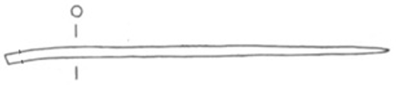
Pin or needle fragment (Fig. 1). The patina is green-blue and granulated, typical of terrestrial context. Dimensions: L = 9cm; Ø = 2.5-2.9mm; WT = 3.6g.
Pin
Hauterive - Champréveyres, Neuchâtel, Neuchâtel, Switzerland
Excavation 1983-1985, object from layer 1 (containing material from the Bronze Age until the 20th century)
Late Bronze Age
Hallstatt A2/B (1050BC _ 800BC)
Lake
Laténium, Neuchâtel, Neuchâtel
Laténium, Neuchâtel, Neuchâtel
Hr 3031
Not conserved
Nothing to report.
Stratigraphic representation: none.
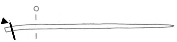
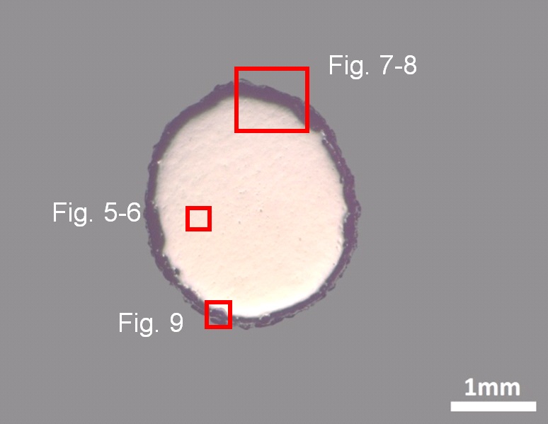
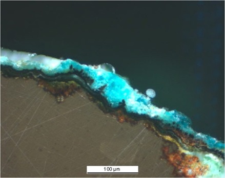
The cross-section is circular and is a complete section through the pin (Fig. 2). The surface is completely covered with a rather thin corrosion crust of irregular thickness (Fig. 3).
Tin Bronze
Cold worked after annealing
MAH 87-195
Musées d'art et d'histoire, Genève, Geneva
Musées d'art et d'histoire, Genève, Geneva
1987, metallography and corrosion characterisation
Nothing to report.
Analyses performed:
Metallography (etched with ferric chloride reagent), Vickers hardness testing, ICP-OES, SEM/EDS, XRD.
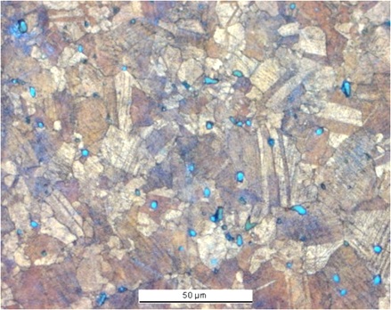
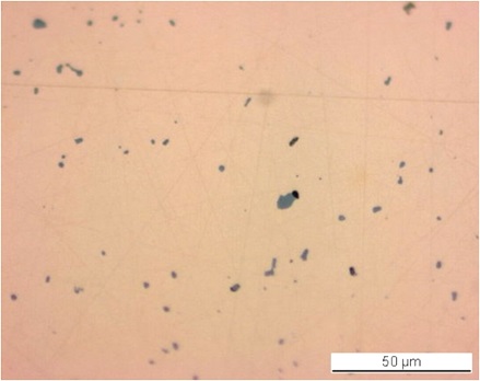
The remaining metal is a tin bronze and contains small copper sulphide and Pb-rich inclusions evenly distributed throughout the metal (Fig. 5, Tables 1 and 2). The Pb-rich inclusions are only visible with SEM appearing as white particles. The etched structure of the tin bronze shows re-crystallised and angular grains, some of them with twins (Fig. 6). Strain or slip lines are visible, especially near the metal surface. They indicate a final cold working. Copper sulphide inclusions are found both at the grain boundaries and inside the grains (Fig. 6). The average hardness of the metal is about HV1 120.
| Elements | Cu | Sn | Sb | Ni | As | Pb | Ag | Co | Zn | Fe |
|---|---|---|---|---|---|---|---|---|---|---|
| mass% | 91.29 | 5.65 | 1.00 | 0.69 | 0.55 | 0.51 | 0.22 | 0.06 | 0.01 | 0.02 |
Table 1: Chemical composition of the metal. Method of analysis: ICP-OES, Laboratory of Analytical Chemistry, Empa.
| Elements | S | Cu | Total |
|---|---|---|---|
| mass% | 21 | 85 | 106 |
Table 2: Chemical composition of grey inclusions (Fig. 5). Method of analysis: SEM/EDS, Laboratory of Analytical Chemistry, Empa.
 Credit HE-Arc CR.
Polygonal and twinned grains + strain lines (metal surface)
Cu
Sn, Sb
Nothing to report.
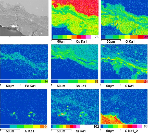
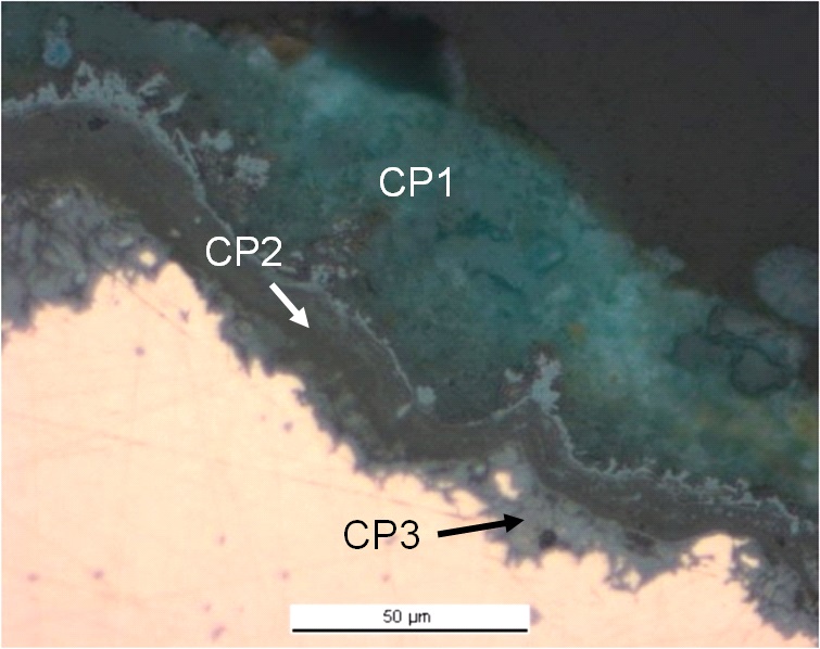
The corrosion crust has an average thickness of about 50µm (Fig. 7). In polarised light (Fig. 8), the corrosion stratigraphy is more clearly visible: it is composed of an inner orange-red corrosion layer (CP3, an agglomerate of nanoscale stannic oxides with cuprite) directly on the metal core (Table 3 and Fig. 9, already studied by Piccardo et al. 2007), an intermediate multi-layered black band (CP2) and an outer turquoise-green layer (CP1) analysed with XRD by Schweizer and identified as malachite/CuCO3Cu(OH)2 (Schweizer 1994, 150). In some areas the orange-red layers (CP3) can also be found in between the black band (CP2) and the malachite (CP1). Elemental chemical distribution of the SEM image of Fig. 9 shows that the black layers are enriched in Sn but also contain Fe (Fig. 9). Superior markers such as contextual Al and Si are present in the outer malachite layers. S is present both on the rim of the outer black layer and in the malachite (Fig. 9, Table 3).
|
Elements |
O | Cu | Sn | S | Cl | Fe | As | Ag | Total |
|---|---|---|---|---|---|---|---|---|---|
| CP3 ext. | 20 | 40 | 12 | 15 | < | 5 | < | 1.9 | 94 |
| CP3 int. | 20 | 53 | 16 | < | 0.9 | < | 0.6 | < | 91 |
Table 3: Chemical composition (mass %) of orange corrosion products (from Figs. 7 and 8). Method of analysis: SEM/EDS, Laboratory of Analytical Chemistry, Empa.
 Credit HE-Arc CR.
 Credit HE-Arc CR.
Multiform - pitting
Type II (Robbiola)
Nothing to report.
Corrected stratigraphic representation: none.
The pin is made from a tin bronze and has been repeatedly cold worked and annealed. After the last annealing there has been some cold work, as can be seen from the strain lines visible after etching the metal. Due to the presence in the corrosion crust of an outer malachite layer, the corrosion was described as terrestrial by Schweizer (Schweizer 1994). The elemental chemical distribution of the corrosion crust shows a more complex situation: as expected for an object buried in a terrestrial site, a typical enrichment of Sn is observed in the inner and intermediate layers covering the remaining metal surface. However it is combined with Fe and S which are often present in lake patinas. According to Schweizer, these layers were formed in anaerobic conditions and developed later on into malachite in an aerated soil through partial dehydration (Schweizer 1994, Schwartz 1934). Since the original surface is absent (destroyed), we refer to type 2 corrosion after Robbiola et al. 1998.
|
References on object and sample |
|
References object 1. Rychner-Faraggi A-M. (1993) Hauterive – Champréveyres 9. Métal et parure au Bronze final. Archéologie neuchâteloise, 17 (Neuchâtel), planche 74.11.
References sample 2. Empa Report 137 695/1991, P.O. Boll.3. Rapport d'examen, Laboratoire Musées d'art et d'Histoire, Geneva GE (1987), 87-194 à 197. 4. Schwartz, G.M. (1934) Paragenesis of oxidised ores of copper, Economic Geology, 29, 55-75. 5. Schweizer, F. (1994) Objets en bronze provenant de sites lacustres: de leur patine à leur biographie. In: L'œuvre d'art sous le regard des sciences (éd. Rinuy, A. and Schweizer, F.), 143-157. |
|
References on analytic methods and interpretation |
| 6. Interpretation of orange corrosion products, see: Piccardo P., Mille B., Robbiola L. Tin and copper oxide in corroded archaeological bronzes, In: Corrosion of metallic heritage artefacts, European Federation of Corrosion Publication n°48, 2007, ed. Dillmann et al, 239-262. 7. Robbiola, L., Blengino, J-M., Fiaud, C. (1998) Morphology and mechanisms of formation of natural patinas on archaeological Cu-Sn alloys, Corrosion Science, 40, 12, 2083-2111. |