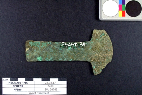
Sacrificial knife (tumi) IVc 24745
Christian. Degrigny (HE-Arc CR, Neuchâtel, Neuchâtel, Switzerland) & Marion. Billot (HE-Arc CR, Neuchâtel, Neuchâtel, Switzerland) & Valentin. Boissonnas (HE-Arc CR, Neuchâtel, Neuchâtel, Switzerland)
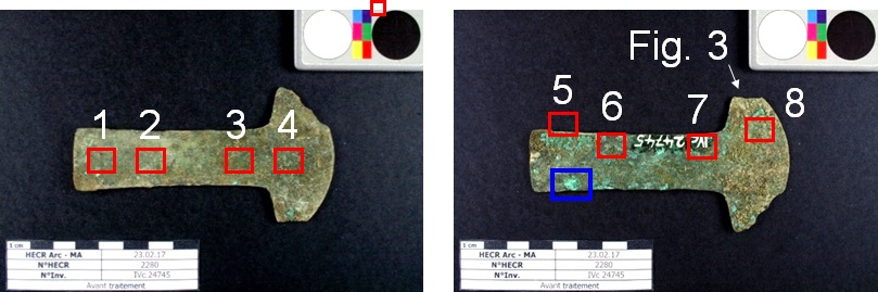
Sacrificial knife. Semi circular flattened cutting edge. Trapezoidal handle with rectangular section. L = 105 mm; W handle = 25 mm; W blade = 55 mm; T = 1 mm; WT = 18,7 g
Sacrificial knife (tumi in vernacular language)
Probably Cajamarca (Peru), Cajamarca, Cajamarca, Peru
Unknown
None
Soil
Museum der Kulturen, Basel
Museum der Kulturen, Basel
IVc 24745
Not conserved
Little information on the origin of the artifact. Most likely not documented before being donated to the museum.
 Credit HE-Arc CR, M.Billot.
 Credit HE-Arc CR, M.Billot.
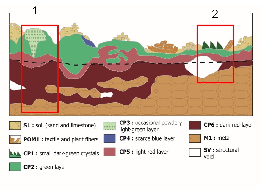
The schematic representation below gives an overview of the corrosion layers encountered on the tumi from a first visual macroscopic observation under binocular microscope.
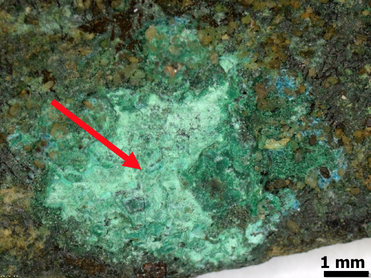
Not applicable since invasive sampling was not authorized by the museum. Only one powder sample was taken of the green corrosion product (Fig. 4).
Cu Alloy
Cold worked (laminated)
1
HE-Arc CR, Neuchâtel, Neuchâtel
HE-Arc CR, Neuchâtel, Neuchâtel
02.04.2017, chemical and structural analyses
Nothing to report.
Analyses performed:
X-ray radiography*, XRF**, SEM-EDS, and XRD. Conditions of the XRD analysis: Stoe Mark II-Imaging Plate Diffractometer System (Stoe & Cie, 2015) equipped with a graphite-monochromator. Data collection was performed using Mo-Kα radiation (λ = 0.71073Å, beam diameter 0.5mm). Two-dimensional diffraction images (10min. per exposure) were obtained at an image plate distance of 200mm with a continued sample rotation. The resolution was Dmin - Dmax 24.00 - 1.04Å and intensity integration has been performed over the entire image (360°).
* The conditions are unknown.
** On the object with portable X-ray fluorescence spectrometer (NITON XL3t 950 Air GOLDD+ analyser, Thermo Fischer®.
The metal is an arsenical copper alloy with low concentration of iron and traces of silver (Table 1). XRF analyses have been conducted on eight areas, on the two sides of the non-cleaned artifact. Almost all analyses are the same except for the area “5” where a slightly higher concentration of arsenic was measured.
|
Elements mass% |
Cu |
As |
Ag |
Fe |
Cl |
Al |
Si |
K |
P |
Ca |
S |
BAL |
|
1 |
61.9 |
0.5 |
0.05 |
0.4 |
0.3 |
3.6 |
12.6 |
0.7 |
0.2 |
1.1 |
0.2 |
18.3 |
|
2 |
51.9 |
0.3 |
0.04 |
0.4 |
0.3 |
1.4 |
11.9 |
0.7 |
0.07 |
0.8 |
0.1 |
31.8 |
|
3 |
51.4 |
0.3 |
0.04 |
0.5 |
0.3 |
2.5 |
14.4 |
0.8 |
0.1 |
0.9 |
0.1 |
28.2 |
|
4 |
52.7 |
0.3 |
0.04 |
0.5 |
0.4 |
2.6 |
16.6 |
0.8 |
0.06 |
1.1 |
0.2 |
24.5 |
|
5 |
39.8 |
1.1 |
0.03 |
0.9 |
2.2 |
3.1 |
13.6 |
0.8 |
0.07 |
1.0 |
0.4 |
36.7 |
|
6 |
52.6 |
0.3 |
0.03 |
0.3 |
0.2 |
1.8 |
11.7 |
0.7 |
0.04 |
1.5 |
0.4 |
30.3 |
|
7 |
52.5 |
0.3 |
0.04 |
0.3 |
0.3 |
2.1 |
11.5 |
0.6 |
0.2 |
1.6 |
0.3 |
30.1 |
|
8 |
38.4 |
0.2 |
0.03 |
0.8 |
0.1 |
3.0 |
16.5 |
0.8 |
0.1 |
0.9 |
0.2 |
38.8 |
None
Cu
As
Complementary information given by X-radiography (Fig. 7) shows that the object is not totally mineralized and still has a lot of remaining metal. Irregularities and different thicknesses are visible and parallel lines show that the knife was hammered. The whiter the area, the thicker the metal is.
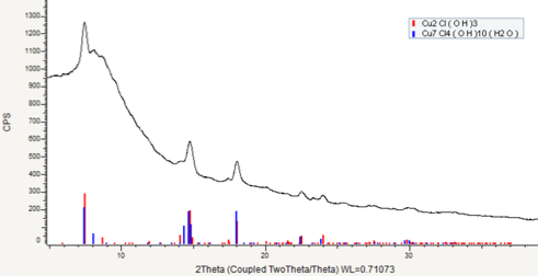
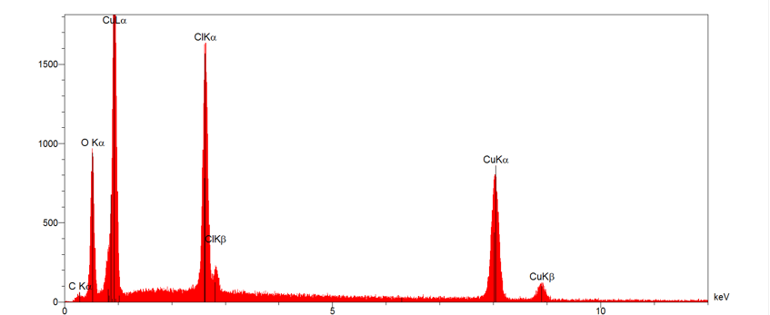
Results from XRF analyses of the non-cleaned metal surface (Table 1) indicate a high concentration of chlorine. The exogenous elements come from the landfill are mainly Si and Al, certainly associated to aluminosilicates. A sample was taken in the powdered light-green isolated layer (Figs. 2, 4 and 5). Chlorine, copper and oxygen was analysed by EDS.SEM. The presence of a high amount of Cl as well as Cu and O seems to indicate active corrosion (Fig. 8). XRD analysis confirmed the presence of paratacamite often associated to active corrosion (Fig. 9).
 Credit HEI-Arc, S.Ramseyer.
Multiform - transgranular
Type II (Robbiola)
Nothing to report.
The schematic representation of corrosion layers of Fig. 5 integrating additional information based on the analyses carried out is given in Fig. 10. The limit of the original surface (represented by the dotted line on the figure below) was identified as still present and is located at the interface between CP6 and CP7.
This tumi is an arsenic copper alloy with a low percentage of iron, as well as traces of silver. This alloy was common in pre-Columbian South America (Pillsbury 2001). The X-ray radiography shows that this object was formed from a metal sheet and cold hammered.
Chlorine is found locally in the form of paratacamite.
The limit of the original surface has been altered by the corrosion products but can be found at the interface of CP6 and CP7. This tumi follows the type II corrosion model of L. Robbiola.
| References on object and sample |
|
References object |
| References on analytic methods and interpretation |
|
2. Scott, D. (2002) Copper and Bronze in Art: Corrosion, Colorants, Conservation. Getty Conservation Institute, Los Angeles. |