Curved pin or tang FK 2670.20
Marianne. Senn (EMPA, Dübendorf, Zurich, Switzerland) & Christian. Degrigny (HE-Arc CR, Neuchâtel, Neuchâtel, Switzerland)
Curved pin or tang (Fig. 1). Several samples were taken. Only one is presented here (Fig. 2). The metal is covered with black corrosion products attributed to a burning process mixed with green corrosion products. Dimensions: L = 114mm; Ø = 4.6mm; WT = 16.7g.
Not defined
Steinmöri, Neftenbach / Dorf Neftenbach, Zurich, Switzerland
Excavation in 1991?, grave 18
Late Bronze Age
14th Century BC, Bronze Age D
Soil
Kantonsarchäologie, Dübendorf, Zurich
Kantonsarchäologie, Dübendorf, Zurich
FK 2670.20
Not conserved
Nothing to report.
Stratigraphic representation: none.
The sample (Fig. 3) is a section from the pin (or tang). The metal is covered by a thick corrosion crust (blue layer adhering to the metal, topped by a green layer mixed with black corrosion products - Museum report (1992)). Dimensions: L = 1.8mm; W = 1.2mm.
Tin Bronze
Secondary recrystallization (produced by burning)
MAH 92-5-4-002
Musées d'art et d'histoire, Genève, Geneva
Musées d'art et d'histoire, Genève, Geneva
1992, examination of metallography examination
Nothing to report.
Analyses performed:
Metallography (etched with ferric chloride reagent), Vickers hardness testing, EPMA/WDS, SEM/EDS.
The remaining metal is a tin bronze (Table 1) with copper sulphide inclusions that contain some Fe (Figs. 5 and 6, Table 2). After etching, the tin bronze shows polygonal grains with few twins (Fig. 6). The grain size varies between 70 and 180µm indicating grain growth due to an extended or excessively hot annealing process. The copper sulphide inclusions appear in blue. The average hardness of the metal is HV1 70.
| Elements | Cu | Sn | Pb | As | Sb | Fe | Zn | Ag | Au | Co | Bi | Ni | S |
|---|---|---|---|---|---|---|---|---|---|---|---|---|---|
| mass% | 89.94 | 8.11 | 0.63 | 0.41 | 0.29 | 0.24 | 0.13 | 0.1 | 0.1 | 0.04 | 0.01 | < | n. d. |
Table 1: Chemical composition of the metal. Method of analysis: EPMA/WDS, Lab Department of Materials, University of Oxford.
|
Elements |
S | Fe | Cu | Total |
|---|---|---|---|---|
| Dark-blue inclusion | 21 | 2.8 | 77 | 102 |
Table 2: Chemical composition (mass %) of the dark-blue inclusions on Fig. 4. Method of analysis: SEM/EDS, Laboratory of Analytical Chemistry, Empa.
 Credit HE-Arc CR.
Large polygonal grains with few twins
Cu
Co, Zn, As, Ag, Sn, Sb, Pb, Fe
Nothing to report.
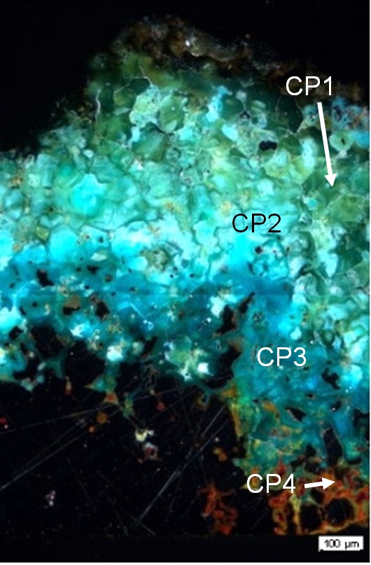
The corrosion crust has an average thickness of 500µm but can in areas be much thicker (Fig. 3). It is divided in two layers. The inner layer is itself divided in two sub-layers: a thin light-grey sub-layer at the interface with the remaining metal surface (CP4, in bright field) which appears red-orange in polarised light (Fig. 7) topped by a medium-grey sub-layer (in bright field) that contains remnant metal (CP3). It turns dark-blue in polarised light (Fig. 7). The outer corrosion layer can also be divided into two sub-layers: a porous sub-layer (CP2) followed by a dark-grey sub-layer in which crystals are outlined by cracks (CP1, in bright field). Under polarized light, the latter turns blue-green while on top it appears olive and brown (Fig. 7). Chemically the corrosion crust is Sn enriched and Cu-depleted (Table 3, Figs. 8 and 9). The maximum of the Sn enrichment occurs on the outer olive and brown sub-layer (CP2). The corrosion layer also contains P, Si, C and O. Inclusions containing Fe or Ag can be found in the corrosion crust (Figs. 8 and 9).
|
Elements |
O | Cu | Sn | Si | P | Fe | Pb | S | Cl | Total |
|---|---|---|---|---|---|---|---|---|---|---|
| CP1, outer corrosion layer | 27 | 17 | 57 | 1.1 | 1.9 | 0.9 | 1.0 | < | < | 105 |
| CP3, inner corrosion layer | 39 | 27 | 26 | 1.1 | 2.5 | < | < | < | < | 97 |
| Remnant metal in CP3 | < | 88 | 8.3 | < | < | < | < | < | < | 97 |
Table 3: Chemical composition (mass %) of the corrosion crust from Fig. 7. Method of analysis: SEM/EDS, Laboratory of Analytical Chemistry, Empa.
 Credit HE-Arc CR.
Fig. 7: Micrograph of the metal sample from Fig. 3 (detail) and corresponding to the stratigraphy of Fig. 4, unetched, polarised light. At the metal - corrosion products interface the colour of the corrosion layer is dark-blue, changing to green-blue in the outer part. The top surface of the corrosion crust is olive to brown,
 Credit Empa.
Fig. 8: SEM image, BSE-mode, and elemental chemical distribution of the selected area from Fig. 3 (due to repolishing before SEM/EDX investigation, the SEM image is slightly different from the area indicated in Fig. 3). The rectangle in the SEM image marks the detail mapping of Fig 9. Method of examination: SEM/EDS, Laboratory of Analytical Chemistry, Empa,
Uniform - intergranular
Type II (Robbiola)
Nothing to report.
Corrected stratigraphic representation: none.
The tin bronze was exposed to an extended or excessively hot annealing process. This, combined with the extreme thickness of the corrosion crust and the dark surface, confirms that the object originates from a fire burial context. At the metal - corrosion crust interface some copper oxide (cuprite?) occurs. On top copper carbonates (azurite or malachite?) are mixed with tin oxide (cassiterite/SnO2?). Tin oxide dominates in the brown-black extremely Sn-rich outer layer. The P-enrichment in the whole corrosion layer may be due to an environment rich in organic material (for example bones). The original surface of the metal has been destroyed, presenting a type 2 corrosion layer after Robbiola et al. 1998.
|
References on object and sample |
|
Reference object Reference sample |
|
References on analytic methods and interpretation |
| 4. Robbiola, L., Blengino, J-M., Fiaud, C. (1998) Morphology and mechanisms of formation of natural patinas on archaeological Cu-Sn alloys, Corrosion Science, 40, 12, 2083-2111. |


