Oval bracelet with a rounded diameter B B3474
Marianne. Senn (Empa, Dübendorf, Zurich, Switzerland) & Christian. Degrigny (HE-Arc CR, Neuchâtel, Neuchâtel, Switzerland)
Bracelet with a rounded diameter after Paszthory (1985, 243). Ends shaped to form little paws, with a casting seam on the inside (Fig. 1). Dimensions: Øobject = around 6.2cm.
Jewellery
Les Eaux-Vives, Genève, Geneva, Switzerland
Unknown
Late Bronze Age
Hallstatt B2/3 (1000BC _ not defined)
Lake
Musées d'art et d'histoire, Genève, Geneva
Musées d'art et d'histoire, Genève, Geneva
B B3474
N/A
None.
None.
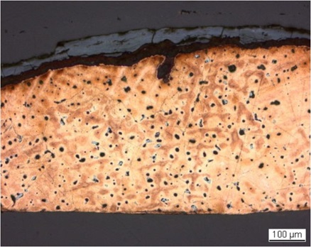
The sample is a section from one end of the bracelet (Fig. 2). Its dimensions are: L = 2.3mm and W = 0.55mm. The corrosion layer is relatively thin (Fig. 3).
Leaded Bronze
As-cast
MAH 77-110-2
Musées d'art et d'histoire, Genève, Geneva
Musées d'art et d'histoire, Genève, Geneva
1977 and 1991, study of the corrosion layer, metal composition
None.
Analyses performed:
Metallography (etched with ferric chloride reagent), Vickers hardness testing, ICP-OES, SEM/EDS.
None.
The remaining metal is a porous leaded bronze (Table 1). Under bright field light Pb and dark-grey copper sulphide inclusions can be seen (Fig. 4, Table 2). The copper sulphide inclusions are rather small with various forms, while the Pb inclusions are generally larger and round. The etched leaded bronze has the dendritic structure of an as-cast metal (Fig. 5) with an average hardness of HV1 80. After etching the Pb-inclusions turned dark grey and the copper sulphide light grey (Fig. 5).
| Elements | Cu | Sn | Pb | Sb | As | Ni | Ag | Co | Zn | Fe | Bi |
|---|---|---|---|---|---|---|---|---|---|---|---|
| mass% | 89.67 | 6.40 | 2.62 | 0.52 | 0.27 | 0.22 | 0.13 | 0.07 | 0.03 | 0.04 | 0.03 |
Table 1: Chemical composition of the metal. Method of analysis: ICP-OES, Laboratory of Analytical Chemistry, Empa.
|
Elements |
S | Cu | Total |
|---|---|---|---|
| Dark-grey inclusion | 21 | 76 | 97 |
Table 2: Chemical composition (mass %) of dark-grey inclusions on Fig. 4. Method of analysis: SEM/EDS, Laboratory of Analytical Chemistry, Empa.
 Credit HE-Arc CR.
Dentritic structure with pores and inclusions
Cu
Ni, As, Ag, Sn, Sb, Pb
None.
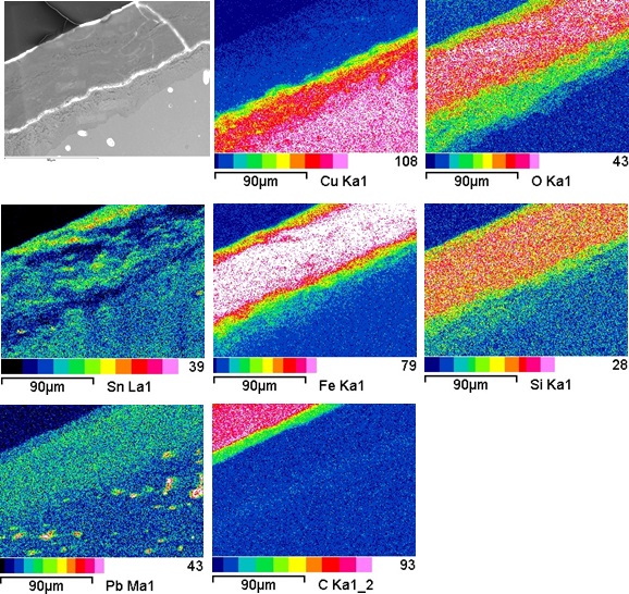
The corrosion crust has an average thickness of 60µm and is composed of two main layers (CP2 and CP3, Fig. 4). In bright field, the inner layer (CP3) has a slight blue hue (Fig. 4). The corrosion products are stratified and the outer part is porous (Fig. 6). In polarised light, the inner layer appears as a mixture of reddish and orange corrosion products (Fig. 8). This layer is Cu- and O-rich with some Sn, Fe and Si in the porous zone (Table 3, Fig. 8). In bright field, the outer cracked and stratified layer (CP2) is dark grey (Fig. 5). It is depleted of Cu and rich in Fe and Sn with significant amounts of O and Si (Fig. 8). In polarised light, large red angular crystals (possibly cuprite) appear clearly in this outer corrosion layer (Fig. 7). The surface of the outermost layer (CP1) is Sn and O and Fe enriched (CP1, Fig. 8).
|
Elements |
O | Fe | Cu | Sn | Pb | Si | S | As | Total |
|---|---|---|---|---|---|---|---|---|---|
| CP2 | 40 | 30 | < | 25 | 3.1 | 5 | < | 0.6 | 104 |
| CP3 | 33 | 42 | 3.7 | 8.8 | 2.6 | 4.4 | < | 0.6 | 95 |
| Red angular crystals in CP2 | 17 | 4.3 | 69 | 9.9 | 0.8 | 1.5 | < | < | 103 |
Table 3: Chemical composition (mass %) of the corrosion layers from Figs. 7 and 8. Method of analysis: SEM/EDS, Laboratory of Analytical Chemistry, Empa.
 Credit HE-Arc CR.
 Credit HE-Arc CR.
Fig. 7: Micrograph similar to Fig. 4 and corresponding to the stratigraphy of Fig. 9, polarised light. From bottom left to top right: the metal (in brown with white porosities and blue Pb inclusions), the inner layer (CP3) appearing as stratified red and orange layers followed by an outer layer (CP2, black with red angular crystals) and a superimposed light green layer (CP1),
Uniform - pitting
Type I (Robbiola)
None.
None.
The leaded bronze artefact shows an as-cast structure. The outer layer is typical for a lake patina (though in this case formed under aerobic conditions), containing principally Fe as well as other contextual elements (such as Si). The absence of Cu in the corrosion layer could be due to its re-crystallisation in large cuprite crystals. Surprisingly the top of the outer layer is enriched in Sn, which was not the case in the study carried out by Schweizer (Schweizer 1994). The additional presence of C in this top layer could indicate a secondary, terrestrial patina formation phase. The corrosion is a type 1 according to Robbiola et al. 1998.
References on object and sample
Reference object
1. Paszthory, K. (1985) Der bronzezeitliche Arm- und Beinschmuck in der Schweiz. PrähistorischeBronzefunde X-Bd. 3, München, 243, Tafel 171.
Reference sample
2. Empa report 137'695/1991, P. Boll.
3. Rapport d'examen (1977 and 1991) Laboratoire Musées d'art et d'histoire, Genève (1977-110).
References on analytic methods and interpretation
4. Robbiola, L., Blengino, J-M., Fiaud, C. (1998) Morphology and mechanisms of formation of natural patinas on archaeological Cu-Sn alloys, Corrosion Science, 40, 12, 2083-2111.
5. Schweizer, F. (1994) Objets en bronze provenant de sites lacustre: de leur patine à leur biographie. In: L'œuvre d'art sous le regard des sciences (éd. Rinuy, A. and Schweizer, F.), 143-157.