Deformed fragment of metal sheet WT10-M305
Marianne. Senn (EMPA, Dübendorf, Zurich, Switzerland) & Christian. Degrigny (HE-Arc CR, Neuchâtel, Neuchâtel, Switzerland)
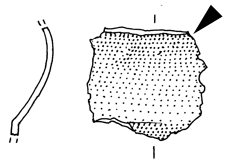
Deformed fragment of metal sheet with a dark green and grey powdery surface (patina) that might have been caused by exposure to high temperatures (Fig. 1). A green layer appears below the dark surface. Dimensions: L = 2.4cm; W = 2.3cm; WT = 4.8g.
Metal sheet
Ritual place Wartau Ochsenberg, Sankt Gallen, Saint Gallen, Switzerland
Excavation in 1991
Iron Age
5th Century BC
Soil
Kantonsarchäologie, Sankt Gallen, Saint Gallen
Kantonsarchäologie, Sankt Gallen, Saint Gallen
WT10-M305
Not conserved
None.
None.
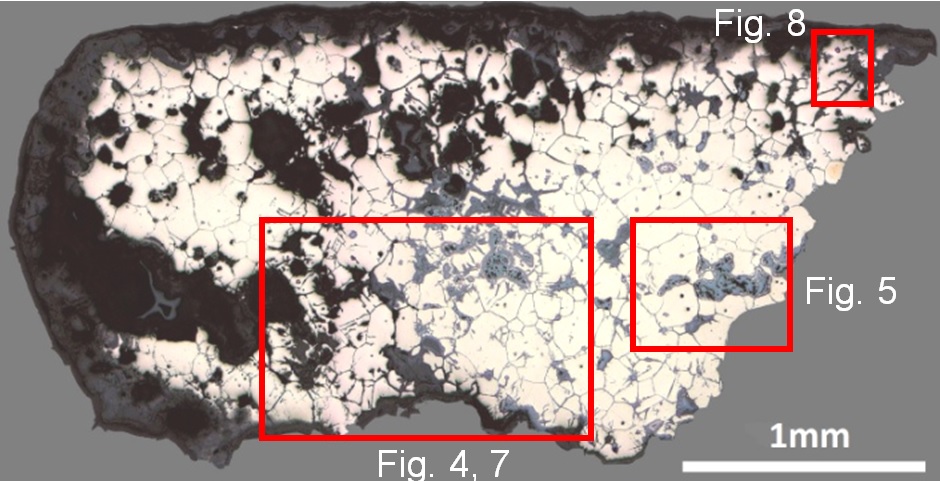
The sample is a section from the top right corner of the sheet (Fig. 2). Its dimensions are: L = 2.5mm and W = 2.3mm. The metal is surrounded on three sides by corrosion products. Intergranular corrosion has developed throughout the metal section (Fig. 3).
Tin Bronze
Secondary recrystallization (produced by burning) after cold working
MAH 92-5-2-003
Musées d'art et d'histoire, Genève, Geneva
Musées d'art et d'histoire, Genève, Geneva
1992, examination of the corrosion layer
None.
Analyses performed:
Metallography (etched with ferric chloride reagent), Vickers hardness testing, ICP-OES, SEM/EDS, Raman spectroscopy.
None.
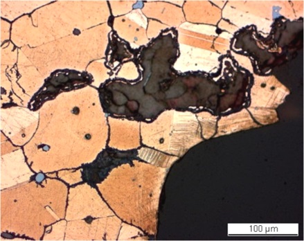
The remaining metal is a porous (red arrows on Fig. 4) tin bronze (Table 1). Five analyses were carried out. S was detected in the non-corroded part of the metal (2 measurements) while P was present only in the corroded metal (3 measurements). As no major difference in the composition was observed (comparison of relative standard deviation, RSD) all analyses were used to calculate the median value. Inter- and transgranular corrosion has developed so extensively that all grain boundaries and twin lines are outlined (Fig. 4). After etching, the metal shows annealed polygonal grains with a few twins and slip lines below the surface (Fig. 5). The slip lines are restricted to the right side of the sample where the metal is best preserved (Fig. 4). The grain size varies between 50 and 170µm, due to an excessively long or hot annealing procedure leading to a grain coarsening. Small copper sulphide inclusions appear in blue (Fig. 4). The average hardness of the metal is HV1 90.
| Elements | Cu | Sn | As | S* | P** | Co | Ni | Pb | Sb | Ag | Zn | Fe | Bi |
|---|---|---|---|---|---|---|---|---|---|---|---|---|---|
| mass% (median value of 5 measurements) | 83.13 | 16 | 0.26 | 0.1 | 0.07 | 0.032 | 0.025 | 0.02 | 0.02 | 0.009 | 0.002 | < | 0.002 |
| RSD % | 2 | 9 | 13 | 25 | 45 | 3 | 5 | 41 | 15 | 12 | 6 | < | 43 |
Table 1: Chemical composition of the metal. Analytical method: LA-ICP-MS, Laboratory of Basic Aspects of Analytical Chemistry at the Faculty of Chemistry, University of Warsaw, PL. *S is only present in the metal, whereas **P indicates the presence of corrosion products in the analysed metal.
 Credit HE-Arc CR
Large polygonal grains with few twins + strain lines
Cu
As, Sn
None.
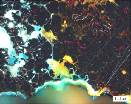
The corrosion crust varies in thickness between 60 and 150µm (Fig. 3). In bright field, it appears dark-grey (Fig. 4) and consists of two layers (CP1 and CP2). The inner layer is dark-grey and dense while the thin outer layer is slightly lighter coloured. Within the metal, the corrosion products are light-grey (CP3, Fig. 4). Under polarized light, the corrosion layer turns blue-green with dark-blue areas (Figs. 6 and 7) whereas corrosion products inside the metal are either light-blue or red-orange (Figs. 6 and 7). The red corrosion products (CP3) have the composition of cuprite/Cu2O while the orange compounds (also CP3) are enriched in Sn (Table 2). The blue-green corrosion products (CP2) both within the remaining metal and on the surface are even richer in Sn and O, and contain some P (Table 2 and Fig. 8). The thin, irregular dark-grey surface layer (CP1) is enriched in P, Fe, Si and Al (Table 2 and Fig. 8). XRD analyses of powdery particles sampled from the thin, dark surface corrosion layer (CP1) indicate the presence of tenorite/CuO and cassiterite/SnO2 (Museum report 1992). The Raman spectra of this layer (Fig. 9) confirmed the presence of tenorite.
|
Elements |
O | Cu | Sn | Si | Fe | P | As | Total |
|---|---|---|---|---|---|---|---|---|
| CP1, outer dark-grey corrosion layer. Fig 7 | 34 | 16 | 49 | < | 3.4 | 3.0 | 0.86 | 108 |
| CP2, blue-green middle corrosion layer. Fig. 7 | 40 | 21 | 41 | 1.4 | < | 1.7 | 0.58 | 106 |
| CP3, Red corrosion product (average of 2 similar analyses). Fig. 7 | 11 | 95 | < | < | < | < | < | 106 |
| CP3, Orange corrosion product (average of 2 similar analyses). Fig. 7 | 24 | 54 | 30 | 0.6 | < | < | 0.59 | 109 |
| Blue-green corrosion product. Fig. 8 | 32 | 21 | 51 | 1.1 | < | < | 1.0 | 106 |
| Blue-green inner corrosion layer. Fig. 8 | 34 | 22 | 39 | 0.8 | < | 1.4 | < | 98 |
Table 2: Chemical composition (mass %) of the different corrosion products and layers from Figs. 6 and 7. Method of analysis: SEM/EDS, Laboratory of Analytical Chemistry, Empa.
 Credit HE-Arc CR.
 Credit HE-Arc CR.
 Credit Empa.
 Credit SNM.
Fig. 9: Raman spectra of the outer dark corrosion layer (S48 and S49) compared to a tenorite standard spectrum. Settings: laser wavelength 532nm, acquisition time 20s for S48 and 100s for S49, one accumulation, filter D1 (7.5-8mW), hole 500, slit 80, grating 600. Method of analysis: Raman spectroscopy, Lab of Swiss National Museum, Affoltern a. Albis ZH,
Uniform - intergranular
Mostly type II with locally type I (Robbiola)
None.
Corrected stratigraphic representation: none.
The tin bronze sheet shows traces of cold working but has been exposed to an extended or excessively hot annealing process. According to Northover (Northover in preparation), the relative lack of twins and their large size confirm a prolonged annealing process. Furthermore large grains, large twins and extensive intergranular corrosion are characteristic of objects that have been exposed to a hot reducing flame either in a house fire or on a funeral pyre. All corrosion products except the cuprite are Sn enriched. The enrichment in P of the surface layer might be due to an environment rich in organic material (for example bones). Tenorite analysed by XRD and Raman spectroscopy is very rare in ancient Cu corrosion and must be interpreted as a further tracer for Cu corrosion in burning context. The original surface of the metal has been destroyed resulting in a type 2 corrosion layer after Robbiola et al. 1998. Only locally in the areas where tenorite is preserved does type 1 patina occur.
References on object and sample
Reference object
1. Publication in preparation (Biljana Schmid-Sikimic).
Reference sample
2. Degli Agosti, M., Santoro, I., Senn, M., Untersuchungen zur Brandpatina an Kupferlegierungen. In: Schmid-Sikimic, B. in preparation.
3. Northover, P. Untersuchungen an Fragmenten einiger Negauer-Helme. In Schmid-Sikimic, B. in preparation.
4. Rapport d'examen (1992) Laboratoire Musées d'art et d'histoire, Genève 92-5-2.
References on analytic methods and interpretation
5. Robbiola, L., Blengino, J-M., Fiaud, C. (1998) Morphology and mechanisms of formation of natural patinas on archaeological Cu-Sn alloys, Corrosion Science, 40, 12, 2083-2111.