Shoe nail 2637 1370.B.
Christian. Degrigny (HE-Arc CR, Neuchâtel, Neuchâtel, Switzerland) & Valentin. Boissonnas (HE-Arc CR, Neuchâtel, Neuchâtel, Switzerland) & Vincent Chappuis. (HE-Arc CR, Neuchâtel, Neuchâtel, Switzerland)
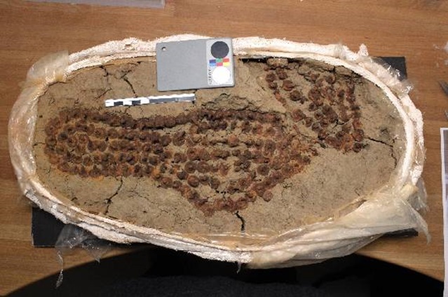
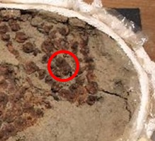
A Roman shoe nail from a block taken from an excavation site (Fig. 1) covered with a thick corrosion crust. The forged nails were hammered into the leather soles on a bucking tool. Average dimensions of the shoe nails: L = about 14mm; diamter = 11mm; W = up to 0.8g.
Shoe nail
Hofstetterfeld, near Roman Necropolis, Sursee, Lucerne, Switzerland
Unknown
Roman Times
2nd half 1st century AD.
Soil
Denkmalpflege und Archäologie des Kantons, Luzern
Denkmalpflege und Archäologie des Kantons, Luzern
2637 1370.B.
Block-lifted (in plaster), desiccated storage with silica gel.
Burial environment: dense sand-clay soil, humid.
The schematic representation below gives an overview of the corrosion layers encountered on the nail from a first visual macroscopic observation.
A longitudinal cut through the nail (Fig. 4). Dimensions: L = 16.4mm; W = about 12.0mm.
Hypoeutectoid steel
Forged
HE-Arc CR 1457-S1.
HE-Arc CR, Neuchâtel, Neuchâtel
HE-Arc CR, Neuchâtel, Neuchâtel
2013, metallography and elementary composition of the metal
None.
Analyses performed:
Metallography (Nital etched), SEM/EDS.
None.
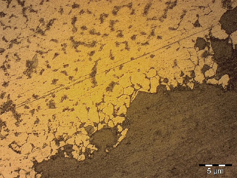
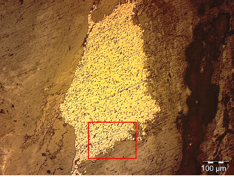
The remaining metal is an hypoeutectoid steel (Fig. 5 and 6), containing Fe, Si and O slags inclusions (possibly fayalite FeO,SiO2), and consisting of less than 1% of the total volume of the highly mineralized nail. After Nital etching, it shows intergranular corrosion on one of its sides (Figs. 7 and 8).
 Credit HEI Arc, S.Ramseyer.
 Credit HEI Arc, S.Ramseyer.
 Credit HEHE-Arc CR, V.Chappuis.
Recrystallized grain structure
Fe
C
None.
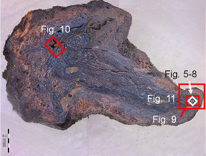
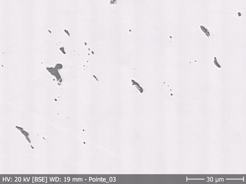
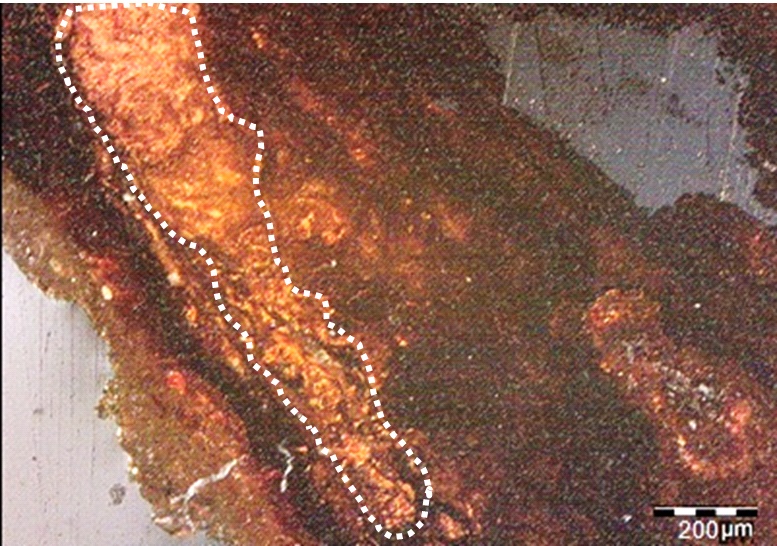
The metal is heavily corroded and the thickness of the corrosion crust is irregular, varying from 0.8 to 3.8mm, with an average of about 2mm. The corrosion has replaced most of the metal (Fig. 4). Between corrosion layers the limit of the original structure is visible. Some leather fibers seem to be preserved as pseudomorphic structures in voids of the metal core (POM1, Figs. 9 and 10). The carbon mapping (Fig.11) shows the presence of carbon in these areas. This can also be due to the penetration of the mounting resin in the cracks.
Si, Al, P and K can be considered external markers, helping to determine the limit of the original surface (Fig. 11 and Table 1). The outer layer of corrosion products (CP1-3) ranges from dark grey to orange in bright field and contains less O and Fe on its borders than the inner layer (CP4-5, Figs. 4 and 11, Table 1). The SEM pictures show a marbling appearance of the inner corrosion layer (CP5). Cl and S do not seem to be present in the sampled nail.
|
Elements Layers |
Fe |
O |
C |
Al |
Si |
K |
P |
|
S1 |
Y- |
Y |
N |
Y++ |
Y+ |
Y |
Y- |
|
CP1 |
Y |
Y |
Y- |
Y |
Y |
Y- |
Y |
|
CP2 |
NA |
NA |
NA |
NA |
NA |
NA |
NA |
|
CP3 |
NA |
NA |
NA |
NA |
NA |
NA |
NA |
|
CP4 |
Y+ |
Y+ |
N |
Y- |
Y- |
N |
Y- |
|
CP5 |
Y+ |
Y+ |
N |
N |
N |
N |
Y-- |
|
NMM |
N |
Y |
N |
N |
Y++ |
N |
NA |
|
POM |
Y+ |
Y- |
Y |
N |
N |
N |
N |
|
SV |
N |
N* |
Y |
Y- |
N |
N |
N |
|
M1 |
Y++ |
N |
N** |
N |
N |
N |
Y-- |
Table 1: Elementary presences in stratigraphic layers based on SEM-EDS elemental mapping (Fig. 13). Y = yes; N = no; NA = not applicable (not observed). + = more compared to other layers; - = less compared to other layers. *Probably pushed back by coating resin. **Though SEM-EDS analysis showed that the metal is an iron-carbon alloy with inclusions of Si, Al, P and K.
 Credit HE-Arc CR, V.Chappuis.
 Credit He-Arc CR, S.Ramseyer.
Uniform - intergranular
aerated soil
None.
The schematic representation of corrosion layers of Fig. 3 integrating additional information based on the analyses carried out is given in Fig. 14. The following strata were identified:
S1 = sand-clay soil, compact, cemented when dry, powdery when abrazed, very thin particle size (usually about and less than 0.1mm). Undefined thickness (soil).
CP1 = powdery orange (loosely coherent) and matte corrosion crust, sometimes forming humps. Partially mixed with S1 and POM layers (diffused interfaces). 1 to 5 millimeters thick.
CP2 = vivid red or pink dots, more brittle than powdery (loose cohesion) and mostly matte, only two or three observed, always above CP3 interfaces. Usually 1 mm spheric diameter.
CP3 = dark reddish corrosion product, forming smooth, matte and brittle layers that are parallel to CP4, with a clean and regular interface. The corrosion products are generally hard, but sometimes break up into grains, resulting in a more irregular interface. The layer is approximately 0.5mm thick.
CP4 = dark grey to black corrosion product, smooth with pseudo-metallic reflection (a bit glossy), very hard, when scratched results in orange powder, with somewhat irregular shape and thickness (ranging from 0.3 to 2mm).
CP5 = dark grey to black corrosion product, generally a bit darker than CP4. Smooth with pseudo-metallic reflection, even harder than CP4 and when scratched it turns into an orange coloured powder. Irregular interface regarding CP4. Clear transition and somewhat parallel from CP4 to CP5. As regards its thickness, it fills the remaining volume.
NMM = silica and calcareous inclusions.
POM = mostly powdery and grain-like material, ranging from orange to dark brown in colour, somewhat glossy and similar to crystals when illuminated. A few fiber-like zones are present, which are more satin than glossy. They are dark brown to charcoal black, very brittle and break into a powdery and grain-like material. POM is located above or close to CP3 and CP4 layers, sometimes mixed into S1 or CP1 layers with a diffused interface. Overall, the stratum is approximately 0.2-0.3mm thick.
SV = structural void, present as fissures that either pass through the different layers, or are present at corrosion interfaces, especially at the CP4/CP5 interface. The void is 0.1 to 0.3mm thick and partially filled with POM in the outer layers.
M1 = glossy with metallic reflection.
The nail is apparently made of a hypoeutectoid steel, with what may be a pearlite phase. Roman shoe nails are forged and pushed through leather soles by hammering them against a hard foot-shaped buckering device. This requires the steel to be strong, but not too brittle, otherwise it will be prone to breaking when pushed through the soles or when in use for marching on hard surfaces. This nail is almost entirely mineralized. Its original shape can be visualised within the corrosion layers. In addition to this, what appears to be leather fibers seem to be preserved in either mineralized or organic form.
References on object and sample
References object
1. Volken, M. (2010) "Le fer et la peau: le cuir et ses outils en milieu urbain romain". In: Chardron-Picault Pascale (dir.). Aspects de l'artisanat en milieu urbain: Gaule et Occident romain. Actes du colloque international d'Autun, 20-22 septembre 2007. Revue Archéologique de l'Est, 28e supplément. RAE, Dijon, 415-424.
2. Volken, M. (2011) Volken Serge et Paccolat Olivier. "Les clous de chaussures du site de Pfyngut: les bases d'une typo-chronologie." In: Paccolat Olivier (dir.). Pfyn/Finges, évolution d'un terroir de la plaine du Rhône. Le site archéologique de "Pfyngut" (Valais, Suisse). Cahiers d'archéologie romande 121, Archaeologia Vallesiana 4, Lausanne, 315-388.