Tang fragment of a knife HR-6246
Marianne. Senn (Empa, Dübendorf, Zurich, Switzerland) & Christian. Degrigny (HE-Arc CR, Neuchâtel, Neuchâtel, Switzerland)
 Credit HEI-Arc CR, N.Gutknecht.
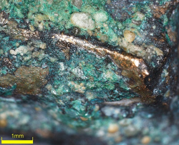
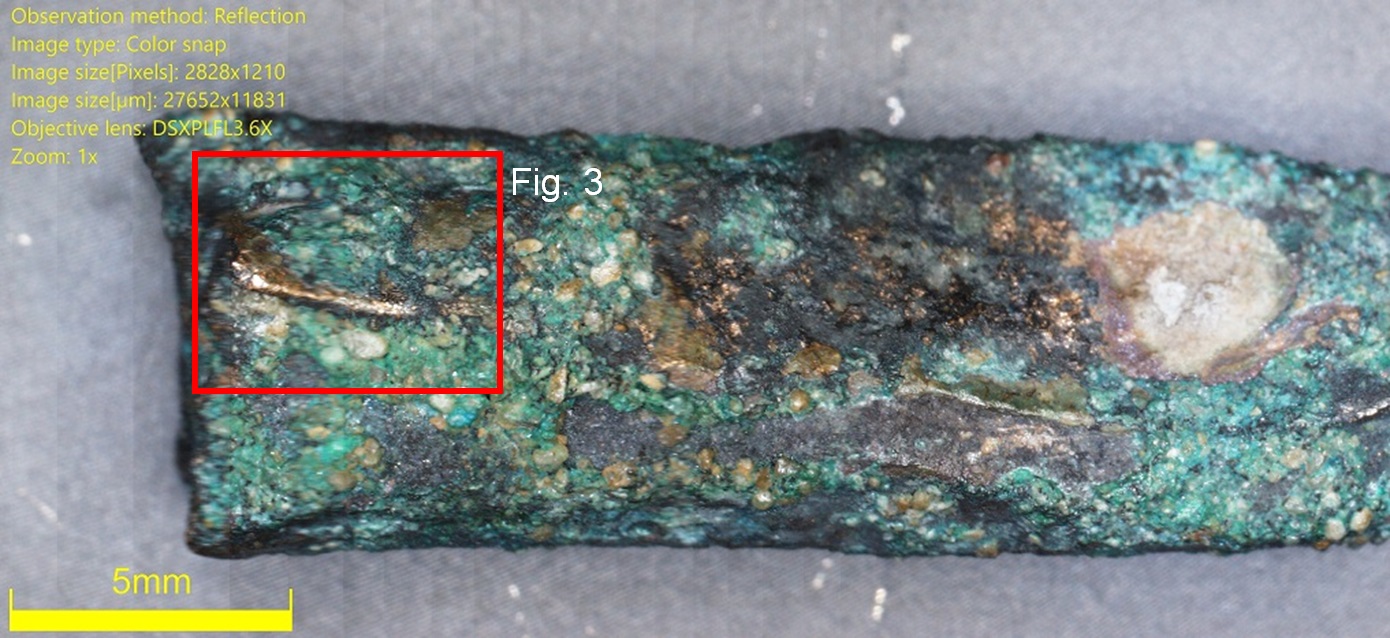
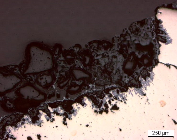
Tang fragment of a knife with lake (shiny brown) and terrestrial (granulated green-blue) crust (Fig. 1). Dimensions: L = 2.7cm; Ø = around 5mm; WT = 5.8g.
Knife
Hauterive - Champréveyres, Neuchâtel, Neuchâtel, Switzerland
Excavation 1983-1985, object from layer 1 (layer with material from Bronze Age till 20th cent.)
Late Bronze Age
Hallstatt A/B
Lake
Laténium, Neuchâtel, Neuchâtel
Laténium, Neuchâtel, Neuchâtel
Hr 6246
N/A
Considered to be a land patina by Schweizer (1994).
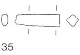
The schematic representation below gives an overview of the corrosion layers encountered on the tang from a first visual macroscopic observation.
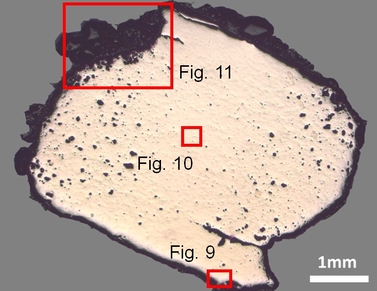
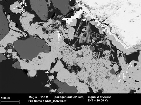
The cross-section corresponds to a lateral cut (Fig. 4). The surface is covered with a thick corrosion crust but part of it has gone (Fig. 8).
Tin Bronze
Cold worked after annealing
MAH 87-197
Musées d'art et d'histoire, Genève, Geneva
Musées d'art et d'histoire, Genève, Geneva
1987, metallography and corrosion characterisation
This sample is mentioned in Schweizer, 1994.
Analyses performed:
Metallography (etched with ferric chloride reagent), Vickers hardness testing, ICP-OES, SEM/EDS, XRD.
None.
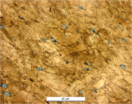
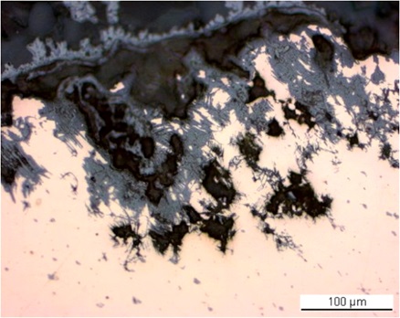
The remaining metal is a tin bronze (Table 1) with high porosity (Fig. 8) and large cracks both on the left and right edges of the sample (Fig. 8). The metal contains small, elongated copper sulphide (Table 2) and Pb inclusions that are oriented parallel to the cracks. Near the metal surface, slip lines are outlined by the development of intergranular corrosion (Fig. 9). The etched metal shows small, elongated grains. Slip lines are also visible (Fig. 10). Annealing is visible in areas near the surface. The average hardness of the metal is HV1 145, but significant variations are observed, depending on where the measurements are taken.
| Elements | Cu | Sn | Sb | Ni | Pb | As | Ag | Co | Fe | Zn |
|---|---|---|---|---|---|---|---|---|---|---|
| mass% | 89.85 | 8.02 | 0.60 | 0.55 | 0.34 | 0.34 | 0.18 | 0.10 | 0.02 | 0.01 |
Table 1: Chemical composition of the metal. Method of analysis: ICP-OES, Laboratory of Analytical Chemistry, Empa.
| Elements | O | S | Cu | Total |
|---|---|---|---|---|
| mass% | 0.9 | 20 | 77 | 98 |
Table 2: Chemical composition of inclusions. Method of analysis: SEM/EDS, Laboratory of Analytical Chemistry, Empa.
 Credit HE-Arc CR.
Elongated grains + strain lines with pores
Cu
Co, Ni, As, Ag, Sn, Sb, Pb
Schweizer (1994) indicates that the copper-tin alloys similar to the one of the tang have minor constituents that were certainly not added intentionally. Furthermore, he mentions that there is no systematic composition difference between bronzes with a lake patina and those with a land patina.
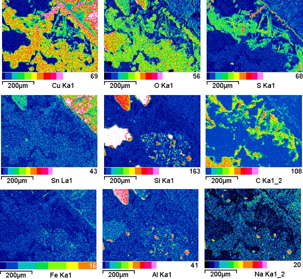
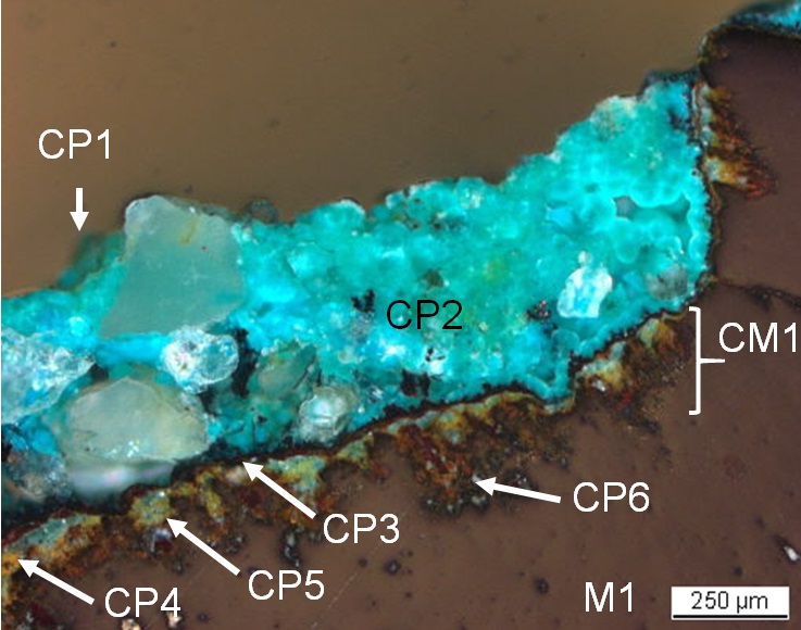
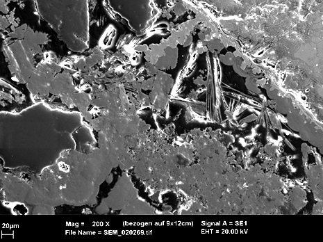
The corrosion crust has a thickness between 0.1mm and 0.7mm (Fig. 8). In bright field (Fig. 11), one can observe that the metal has been replaced by light-grey corrosion products. Adjacent to the metal is a thick, heterogeneous, dark-grey layer with a band of light-grey corrosion products. In polarised light (Fig. 12), all corrosion products which were previously light-grey appear brown-green-yellow, the dark-grey layer being turquoise. The elemental chemical distribution of the SEM images (Figs. 13 and 14) reveals that the inner corrosion products are Sn-rich whereas the adjacent band is Cu and S-rich (Fig. 15). The outer layer contains large inclusions (quartz and others, Si, Al and Na, see Figs. 14-15) and is most probably composed of malachite/Cu2(CO3)(OH)2 (only Cu and O are detected – Fig. 15). S is distributed throughout this layer. XRD analyses indicated the presence of posnjakite/Cu4SO4(OH)6H2O, chalcocite/CuS and djurleïte/Cu1.93S (Schweizer 1994).
 Credit HE-Arc CR.
 Credit HE-Arc CR.
 Credit HE-Arc CR.
Uniform - transgranular
Type I (Robbiola)
None.
Corrosion products CP2 to CP5 observed under binocular corresponds to CP1 to CP4 under cross-section. The other CPs are more difficult to compare, certainly due to the distruption of the corrosion structure during the cutting process.
The tang fragment is made from a bronze and has been cold worked on the top surface after annealing. The past XRD analyses indicate the presence of chalcopyrite in the corrosion crust, typical of lake context (Schweizer 1994), not analysed though with EDX (combinaison of Fe, S and Cu), enriched with Sn close to the metal surface and depleted of Cu on the outer surface. This object was certainly abandoned rather quickly in an anaerobic, humid and S and Fe-rich environment, favouring then the formation of chalcopyrite, before being exposed in an aerated environment in which the crust was formed. The limit of the original surface most probably lies between the Sn-rich inner layer and the Fe and S-rich outer layers. The presence of iron oxides on top of the copper corrosion crust has not yet been explained. The corrosion is a type 1 according Robbiola et al. 1998.
References on object and sample
References object
1. Rychner-Faraggi A-M. (1993) Hauterive – Champréveyres 9. Métal et parure au Bronze final. Archéologie neuchâteloise, 17 (Neuchâtel).
References sample
2. Rapport d'examen, Laboratoire Musées d'art et d'histoire, Geneva GE (1987), 87-194 à 197.
3.Schwartz, G.M. (1934) Paragenesis of oxidised ores of copper, Economic Geology, 29, 55-75.
4. Schweizer, F. (1994) Bronze objects from Lake sites: from patina to bibliography. In: Ancient and historic metals, conservation and scientific research (eds. Scott, D.A., Podany, J. and Considine B.B.), The Getty Conservation Institute, 33-50.
References on analytic methods and interpretation
5. Robbiola, L., Blengino, J-M., Fiaud, C. (1998) Morphology and mechanisms of formation of natural patinas on archaeological Cu-Sn alloys, Corrosion Science, 40, 12, 2083-2111.