Bracelet with dragonflies IIb 4025.01
Perret-Gentil. Emeline (HE-Arc, Neuchâtel, Neuchâtel, Switzerland) & Christian. Degrigny (HE-Arc CR, Neuchâtel, Neuchâtel, Switzerland)
 Credit HE-Arc CR, E.Perret-Gentil.
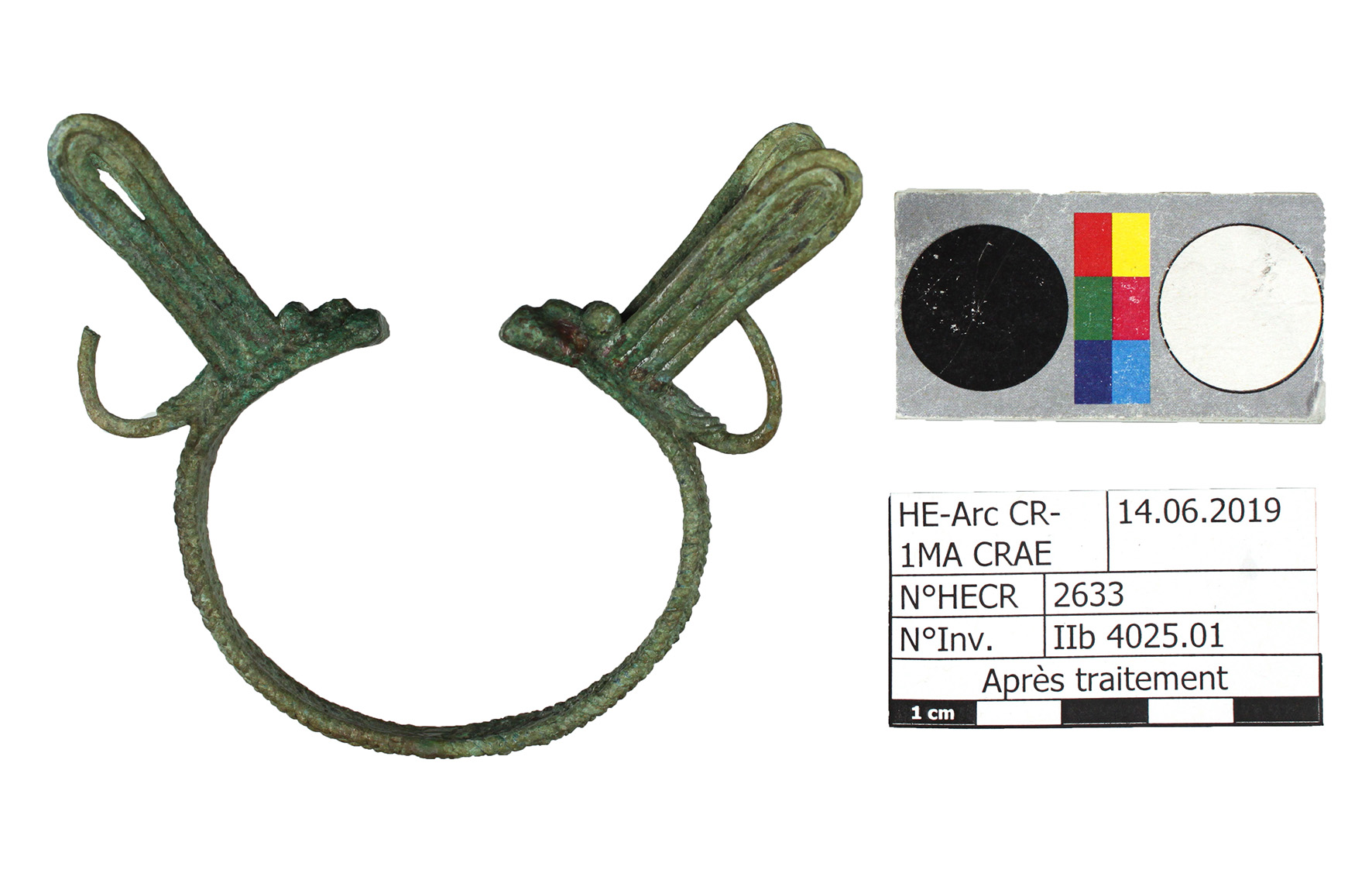
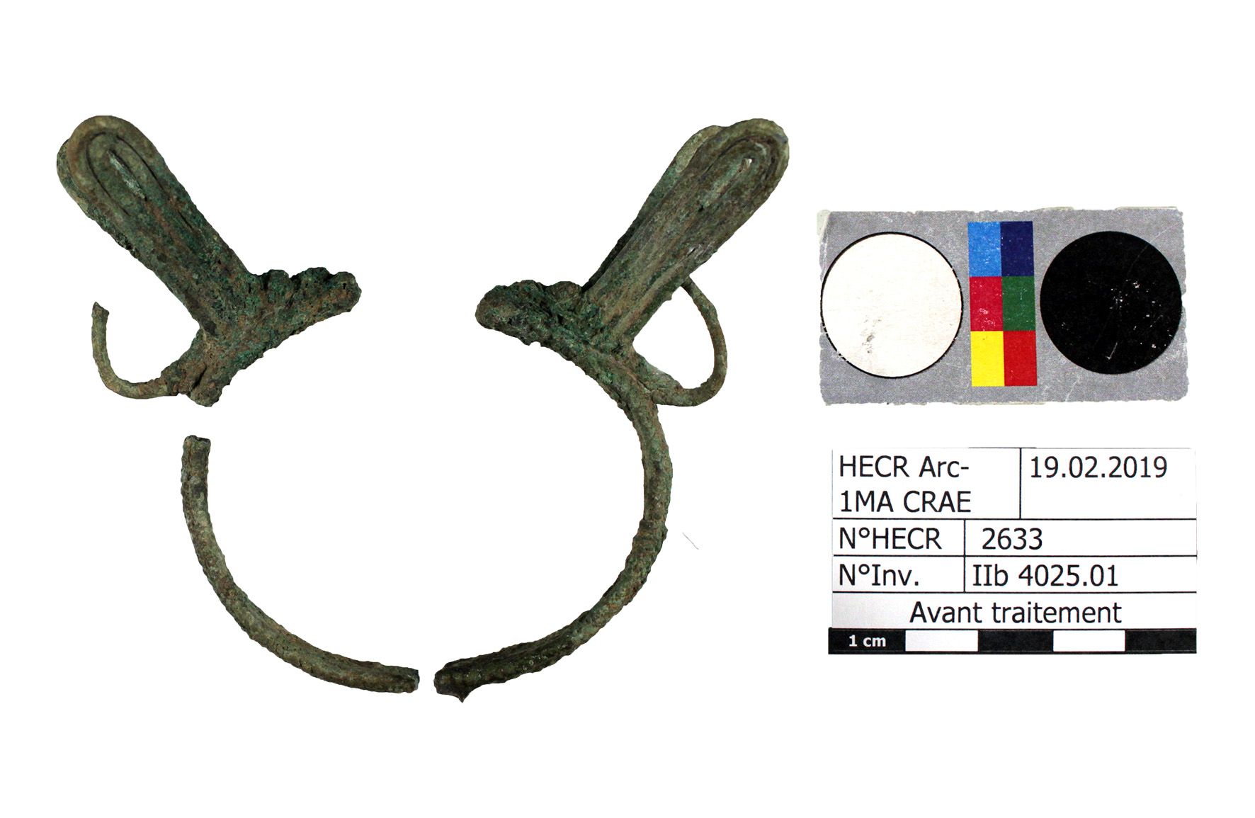
Decorated bracelet with zoomorphic extensions looking like dragonflies (Fig. 1 and 2), covered with dark green corrosion layers and sediments. Dimensions: Ø= 60mm, Lmax = 24mm, WT = 40.82 g (Figs.1 et 2).
Jewellery
Thailand, Udon Thani, Siam, Nong Han, Ban Chiang archaeological site
Date unknown
Late Bronze Age
Soil
Museum der Kulturen, Basel
Museum der Kulturen, Basel
IIb 4025.01
N/A
No information on the archaeological or historical context before 2008 (year of donation to the museum). The artefact was brought to HE-Arc CR in 3 fragments. Preliminary information on corrosion structures could be observed on cross-sections exposed.
 Credit HE-Arc CR, E.Perret-Gentil.
 Credit HE-Arc Ingénierie, S.Ramseyer.
 Credit HE-Arc Ingénierie, S.Ramseyer.
 Credit HE-Arc CR, E.Perret-Gentil.
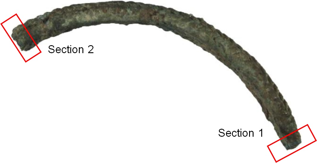
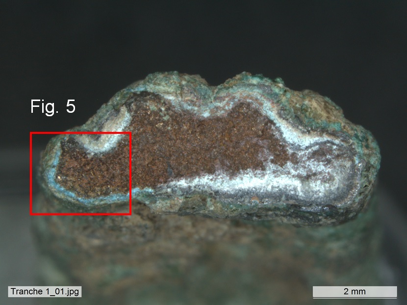
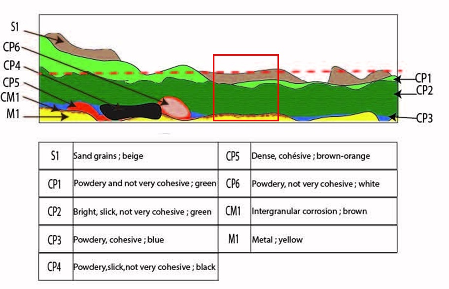
The schematic representation below gives an overview of the strata encountered on the bracelet from a first visual macroscopic observation under a binocular microscope both on the surface and section 2 exposed (Fig. 5).
 Credit HE-Arc CR, E.Perret-Gentil.
 Credit HEI Arc, S.Ramseyer.
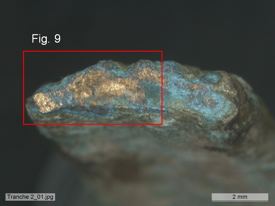
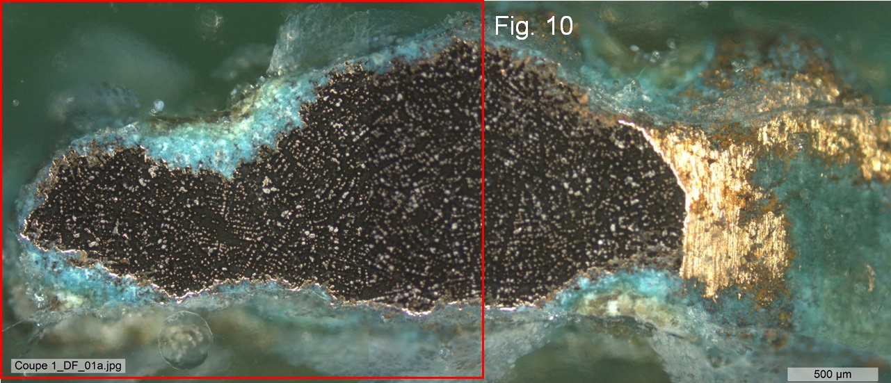
After section 1 of the fragment of Fig. 3 has been consolidated with Technovit 5071 (bicomponent resin (powder+liquid) from Dibenzoylperoxid and Methymethacrylate NN, N-dimethyl-p-tolviol), it was embedded, polished and observed on cross-section (Figs. 9 and 10) to build the MiCorr stratigraphic representation (Fig. 21). Once examined the fragment was extracted, the consolidant was dissolved in acetone and the fragment could be reintegrated on the artefact.
Leaded Bronze
Cast and cold worked
1
Museum der Kulturen, Basel
Museum der Kulturen, Basel
14.05.2019, chemical and structural analyses
Nothing to report.
Analyses performed:
Metallography (etched with ferric chloride reagent), SEM-EDS (Jeol JSM-6400 device), XRD.
None.
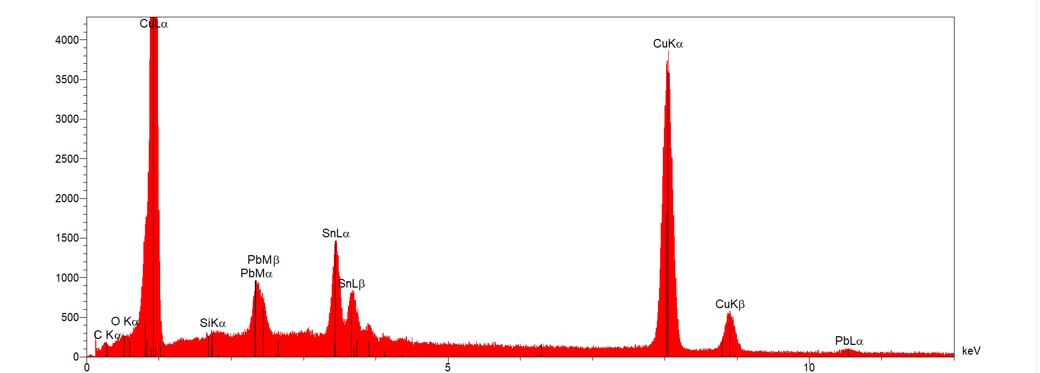
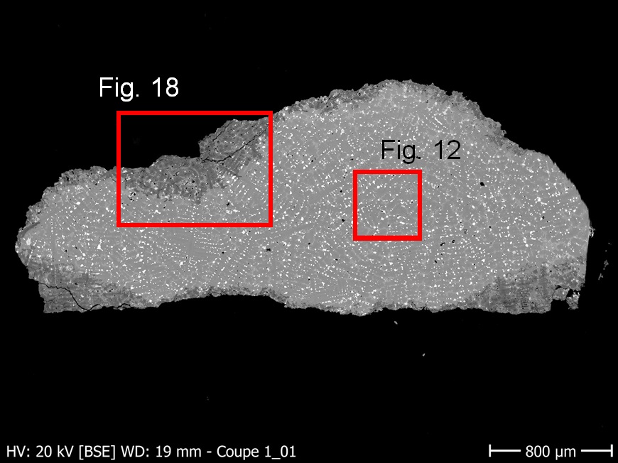
The remaining metal is a porous leaded bronze (Fig. 11) with a high concentration of Sn and Pb (Table 1 and Fig. 12). The 9% Pb concentration is due to the area analysed where Pb nodules false the result which should be more around 5%.
| Elements | Cu | Sn | Pb |
| mass% | 76 | 15-16 | 9 |
Table 1: Chemical composition (mass %) of the metal (from Fig. 12). Method of analysis: SEM/EDS. HE-Arc Ingénierie.
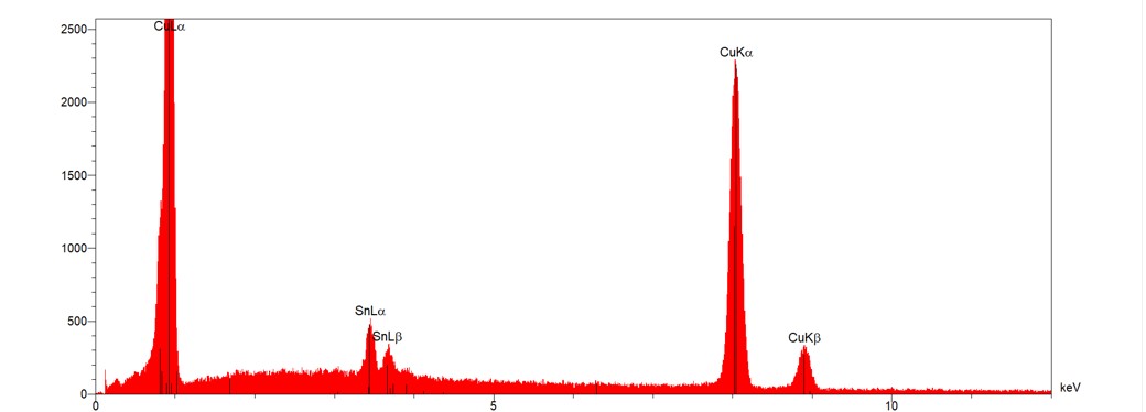
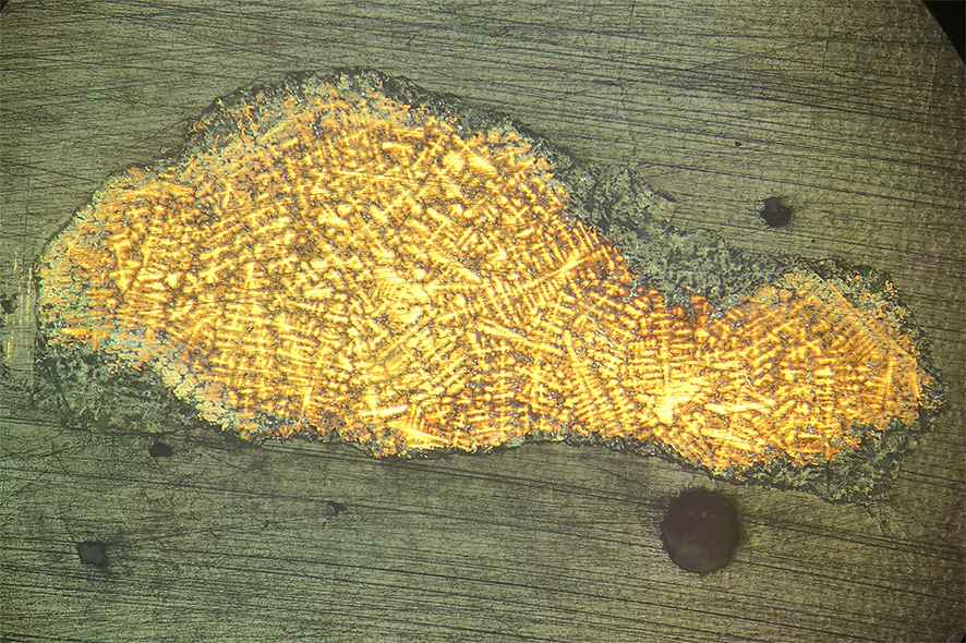
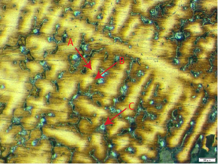
In bright field, the etched alloy shows a dendritic structure (Fig. 13). Therefore, the metal is as-cast. Fig. 14 shows that the yellow inner dendrite phase is richer in Cu (A; Fig. 15) while the orange-brown interdendritic phase is richer in Sn (B; Fig. 16) with lead nodules (C; Fig. 17).
 Credit HE-Arc Ingénierie, S.Ramseyer.
 Credit HE-Arc CR, E.Perret-Gentil.
 Credit HE-Arc CR, E.Perret-Gentil.
Dendritic structure with inclusions
Cu
Sn, Pb
Traces of As have been found locally in some phases of the metal.
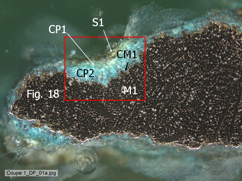
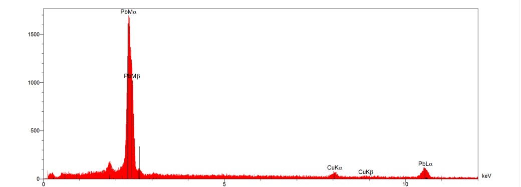
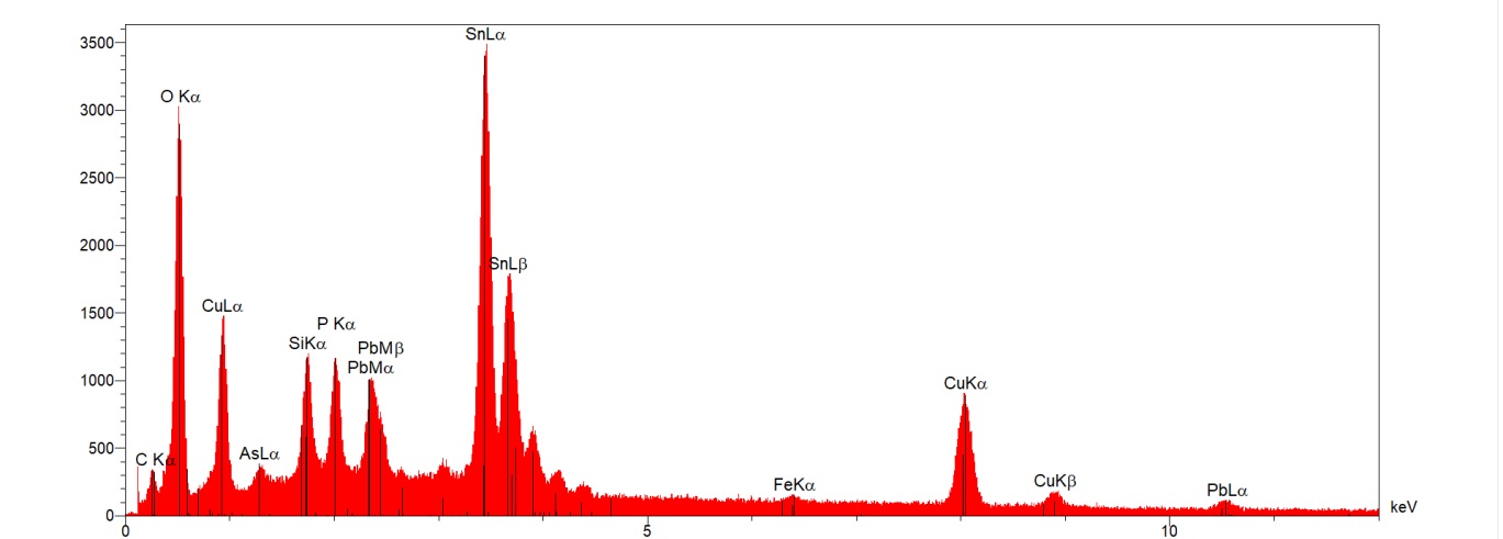
Interdendritic corrosion is visible at the interface metal/corrosion product (Fig. 18) and peudomorph of dendritic structure is visible in CP2 (Figs. 10 and 18). EDS analysis of CP2 and CP1 indicates the presence of higher concentration of Sn in CP2 than CP1 and the presence of P in both layers, validating the funeral context where the object was found. The results of the XRD analysis were irrelevant due to the instability of fragment of Fig. 3 on the support of the device.
 Credit HE-Arc Ingénierie, S.Ramseyer.
Other
Mostly type I with locally type II (Robbiola)
None.
The schematic representation of corrosion layers of Fig. 7 integrating additional information based on the analyses carried out is given in Fig. 22. Strata CP1 and CP2 in the stratigraphy of Fig. 8 are merged into CP1 in Fig. 21. Similarly, strata CP3 to CP6 in Fig. 8 are merged into CP2 and CM1 in Fig. 21.
The metal of the bracelet is a leaded bronze with a high concentration of Sn et Pb. The object was cast and certainly cold-worked to smooth the surface. The metal is heterogeneously corroded with Robbiola types I and II. The limit of the original surface is at the CP1 and CP2 interface. Both layers contain high amounts of Sn and P, validating in the latter case, the funeral context where the object was found.
References on object and samples
1. Scott, D. A. (1991), Metallography and Microstructure of Ancient and Historic Metals. Getty Publications, Los Angeles.
2. Scott, D. A.(2002), Copper and bronze in Art : corrosion, colourants, conservation. Getty Publications, Los Angeles.
3. Rajpitak, W. (1983), The development of copper alloy metallurgy in Thaïland in the pre-buddhist period, with special reference to high-tin bronze, Doctoral thesis , University of London.
4. White, J.C. (2006), Dating Early Bronze at Ban Chiang, Thailand. In From Home erectus to the living traditions: Ühoice of Papers from the 11th International Conference of the European Association of Southeast Asian Archaeologists, Ûougon, 25th-29th September 2006, édité par Pautreau, J.-P, … [et al.], pp. 91-104.