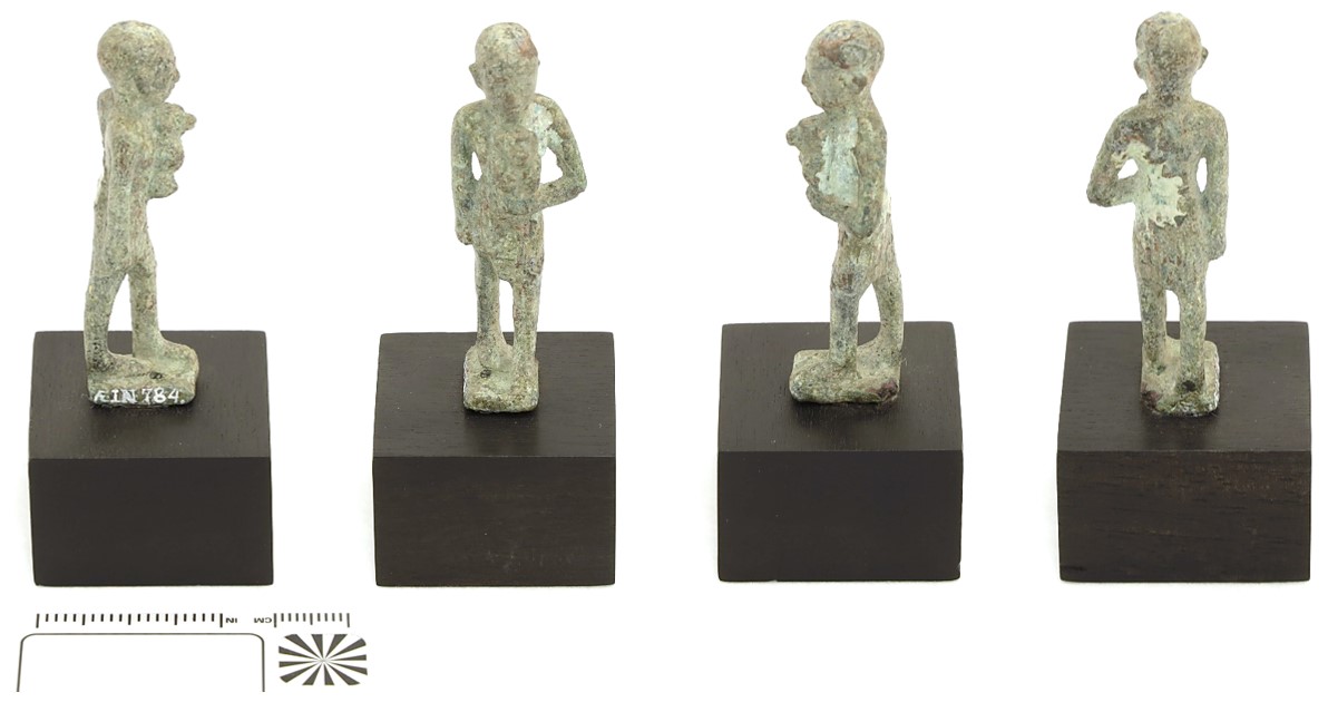
Egyptian figurine of man with baboon - ÆIN 784
Ida. Langemark (The Royal Danish Academy, Copenhagen, Capital Region, Denmark)
The figurine depicts a man carrying a baboon figure, which was probably a sacrifice to the god Thoth. He is wearing a short kilt and a close-fitting hood, and was probably originally part of a composite statuette, where a god, crafted in a much larger scale, was the main motif of the work (Jørgensen, Ægypten V, 2009, p. 282). Height=5.8cm; Width=2.0cm; Thickness=1.8cm.
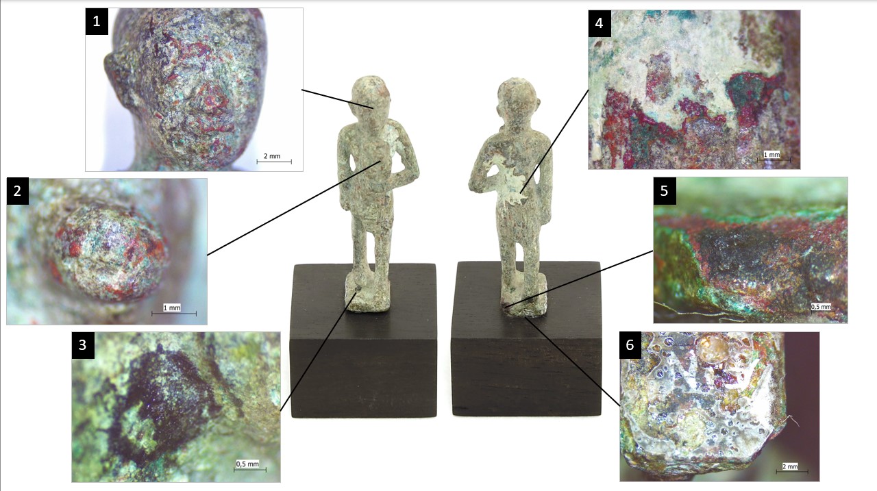
The surface is covered with a white substance with green impurities, located on top of corrosion layers of green, turquoise, red, and dark brown, especially prominent on the figure's back (Fig. 2, picture 4). The proper left corner on the rear side of the figure’s base has been damaged, and the stratigraphy can be easily distinguished (Fig. 2, picture 5).
There are residues of glue under the standing plate (Fig. 2, picture 6).
Votive figure
Archaeological, ancient Egypt, exact provenance unknown
Recovered late 19th century
3rd Intermediate Period to Late Period
None.
Unknown
Ny Carlsberg Glyptotek, Copenhagen
Ny Carlsberg Glyptotek, Copenhagen
ÆIN 784
N/A
Mounted to wooden plinth.
Inventory number applied in white ink of unknown composition.
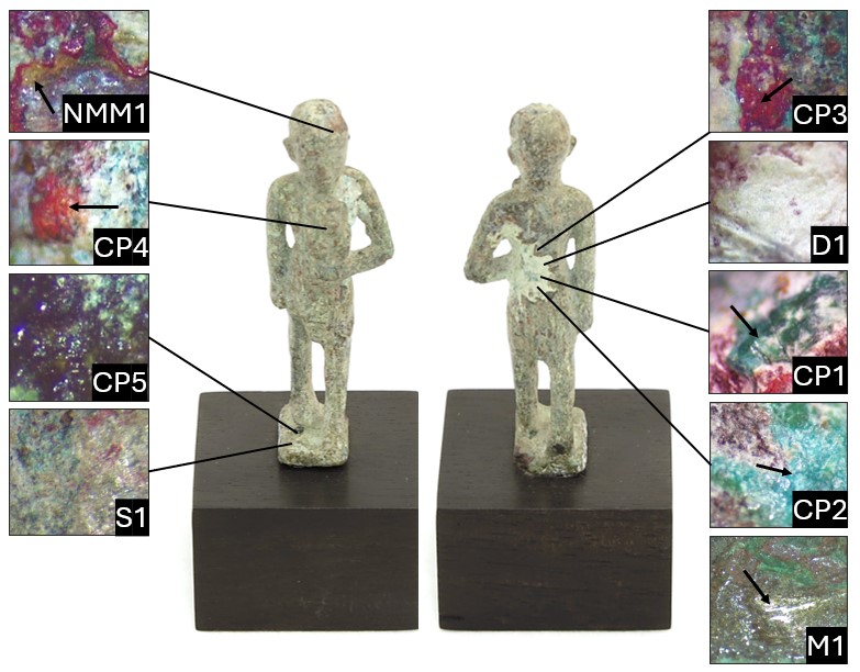 Credit The Royal Danish Academy, I.Langemark.
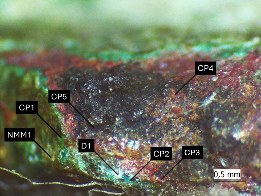 Credit The Royal Danish Academy, I.Langemark.
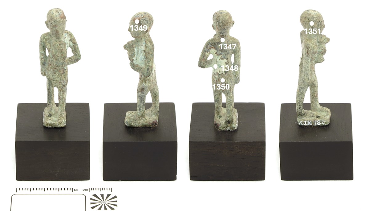
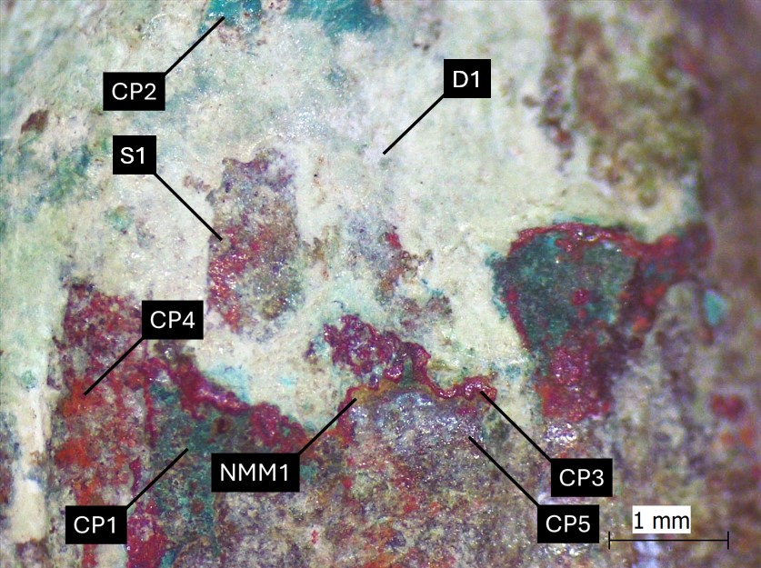 Credit The Royal Danish Academy, I.Langemark.
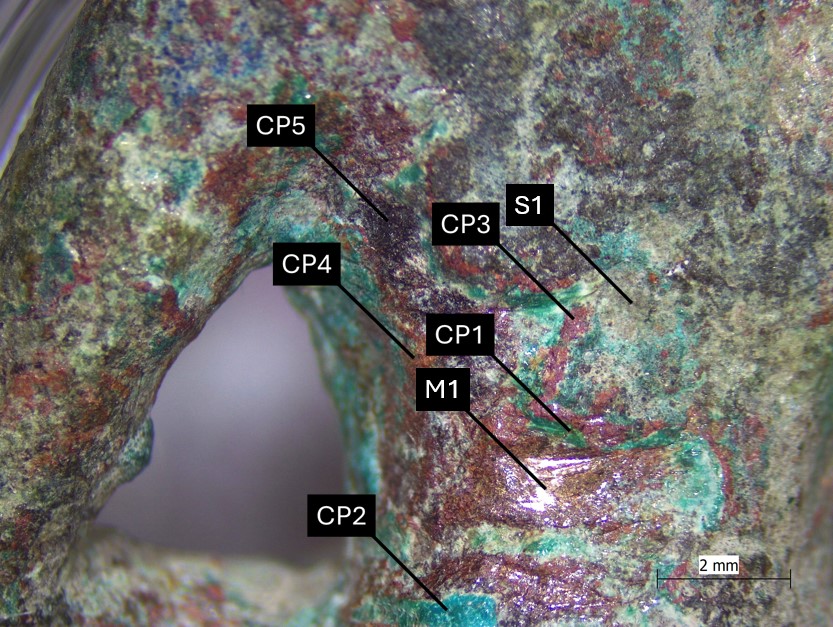 Credit The Royal Danish Academy, I.Langemark.
Stratigraphic representation:
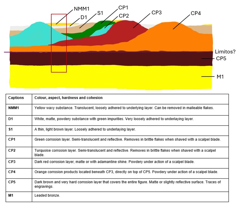
The schematic representation below gives an overview of the corrosion structures encountered on the figurine from a first visual macroscopic observation.
 Credit The Royal Danish Academy, I.Langemark.
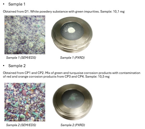
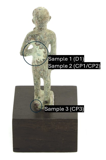
Three samples were taken to be analysed with SEM-EDS and PXRD from areas shown in Fig. 10.
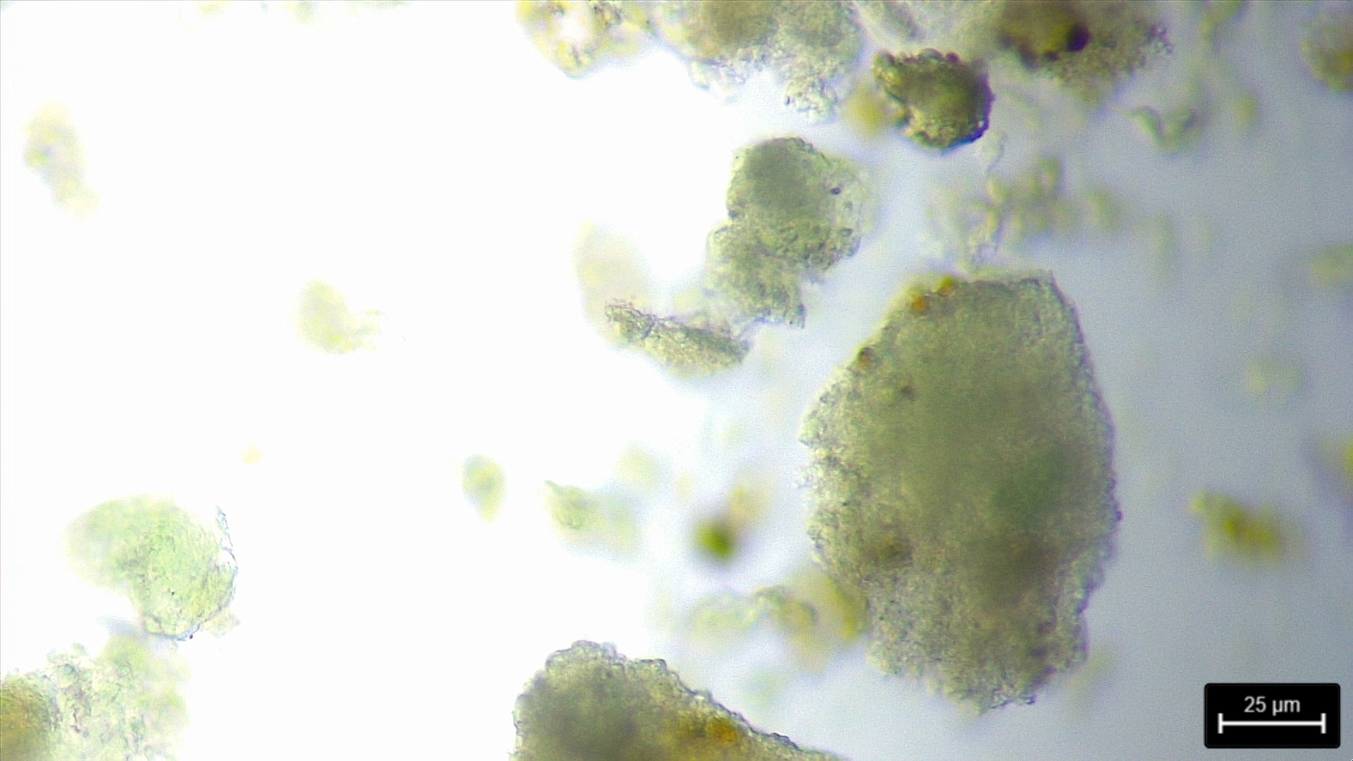
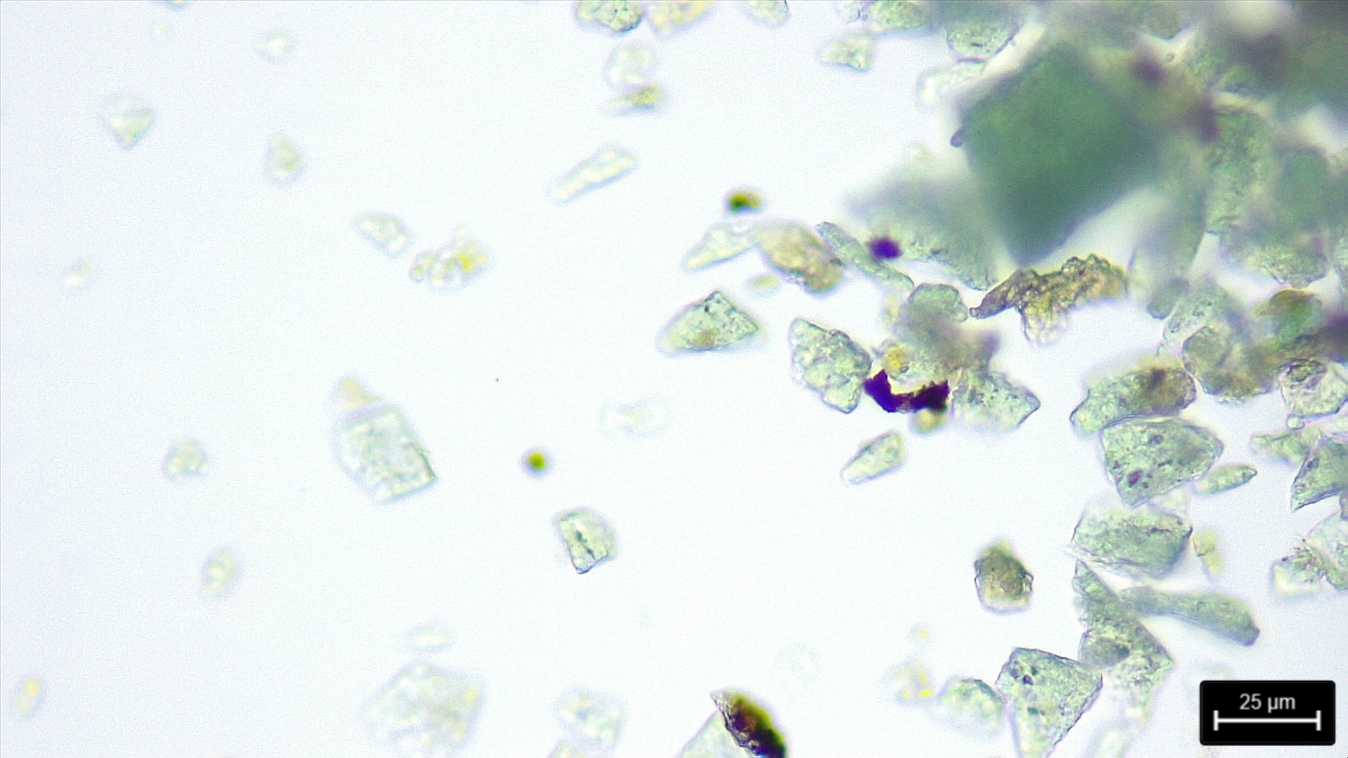
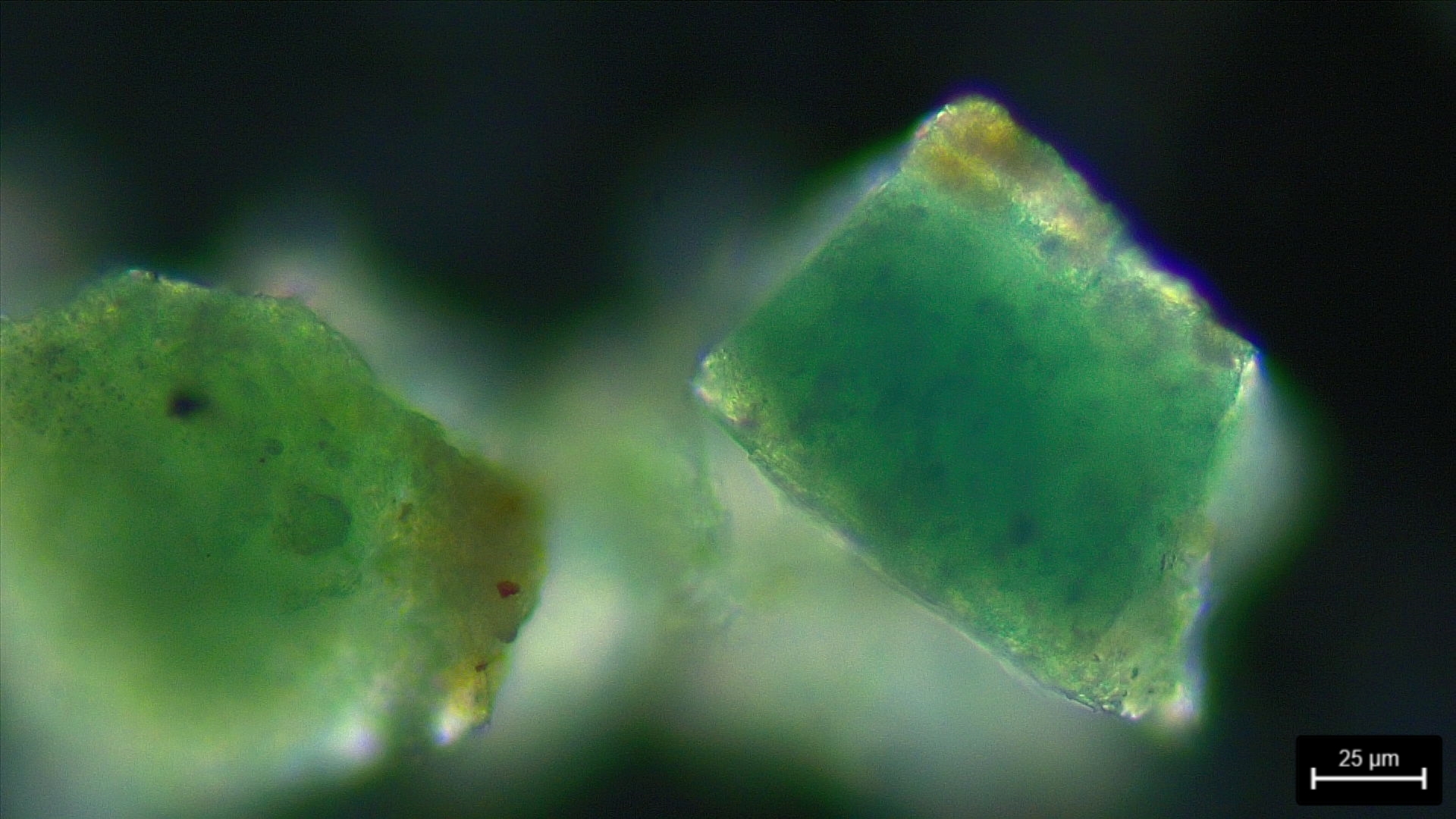
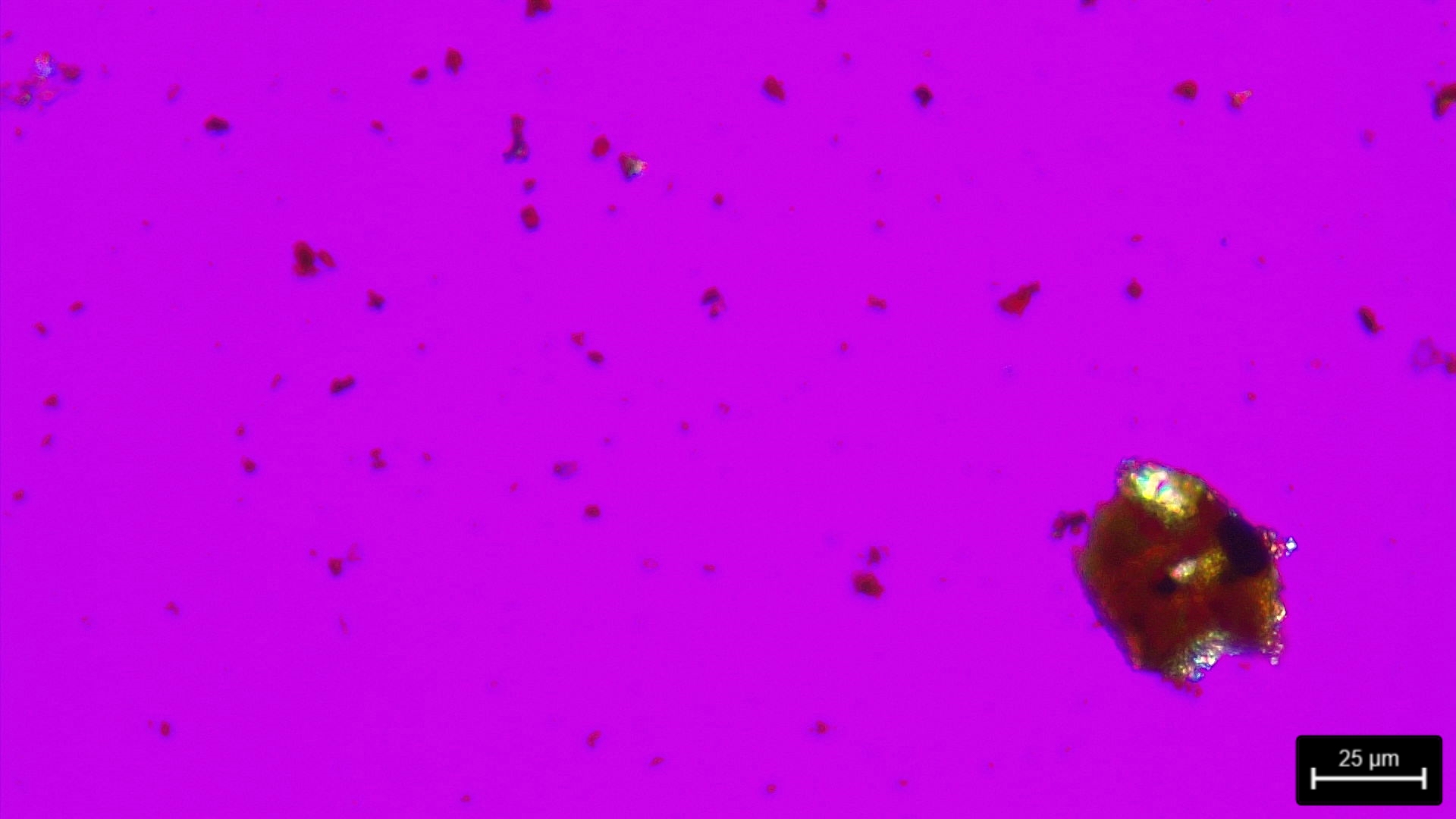
Four samples (1>mg) were observed under polarized microscopy, including material from D1 and C1-C4 (Figures 12-15).
Leaded Bronze
Cast
The Royal Danish Academy (Langemark Ida), Copenhagen
The Royal Danish Academy (Langemark Ida), Copenhagen
January 26th 2024, chemical and molecular analyses of corrosion products.
None.
Analyses performed:
Non-invasive approach
- Portable micro-X-ray fluorescence (μ-XRF) on different measuring locations across the metal surface (Fig. 7). Measurements were made with a portable Bruker Tracer 5G XRF, with an analysis time of 30 sec and a spot size of 8 mm, mode general metal.
Invasive approach
- Polarized microscopy on 4 samples (D1, CP1, CP2, and CP3).
A small amount (1>mg) of material was removed from the surface with scalpel under binocular microscope. The material was placed in a drop of Euparal, covered with an object glass, and left to dry for some days. The samples were observed with a Leica 750P-microscope, and micrographs obtained with the software LAS X.
- Powder X-Ray diffraction (PXRD) of 3 samples. Due to the amount of material required for PXRD-analysis, sampling was done on all areas with a sufficient amount of material, and subsequently grinded into fine powder. PXRD-analyses were performed with a PANalytical X’Pert Pro diffractometer at the Faculty of Pharmacy, Physics and Chemistry, University of Southern Denmark. Measurements were obtained with the software Data collector, and subsequent data processing was done with the applications HighscorePlus and Match!3 respectively.
- Scanning electron microscopy /Energy Dispersive X-ray Spectrometry (SEM-EDS) was performed on 2 samples. The samples were prepared by removing material from the surface with scalpel under binocular microscope. The sample material was attached to steel pins with double sided carbon tape. Analyses were performed with a Hitachi S - 3400N SEM-EDS, 15,0 kV and 300 sec at the Royal Danish Academy, Institute of Conservation.
The μ-XRF-measurements have been performed on unpolished surface areas. They should be seen as indicative. Still the metal is a leaded bronze.
| Measurement no. | Cu | Pb | Sn | Sb | Ag | Zn | Ni |
| 1347 | 48.8 | 45.5 | 4.9 | 0.6 | < | < | < |
| 1348 | 38.9 | 54.2 | 6.0 | 0.8 | < | < | < |
| 1349 | 25.6 | 67.9 | 5.5 | 0.8 | < | < | < |
| 1350 | 34.9 | 56.8 | 7.3 | 0.9 | < | < | < |
| 1351 | 50.3 | 44.9 | 4.1 | 0.6 | < | < | < |
Table 1: Chemical composition (Elements mass (%)) of the figurine in the areas located on Fig. 7.
The μ-XRF-analysis shows a content of approx. 54 % (in weight) lead as an average of measurement 1347-1351. The lead content on the surface of the metal is rather high. The average tin-content is 6,5 %. μ-XRF-measurements show and average of 0,7 % antimony.
None.
none
Cu
Sn, Sb, Pb
None.
The entire metal surface is covered with a dark brown patina, itself covered with various types of corrosion products, mostly Robbiola type I.
Five corrosion products and two non-metallic substances have been identified and can be visualised on Fig. 3 and Fig. 8.
As a reminder:
- NMM1: a yellow waxy substances, presumably remains of coating,
- D1: a white powdery substance with green impurities (Fig. 12),
- S1: a thin light brown layer,
- CP1: a dense green layer (Fig. 13),
- CP2: a dense turquoise layer found at a few localized spots (Fig. 14),
- CP3: a hard red layer (Fig. 15),
- CP4: orange corrosion products located beneath CP4, directly on top of CP5,
- CP5: a dark brown and very hard corrosion layer, that contains corresponding markers to the limit of the original surface.
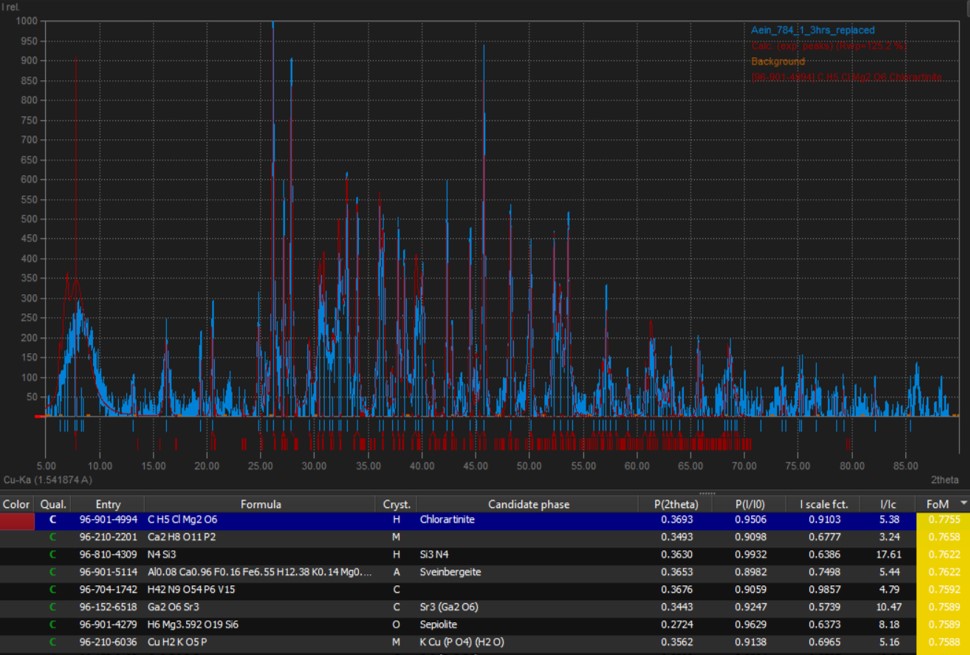
Sample 1, consisting of material obtained from D1, shows under PXRD-analysis the presence of chlorartinite Mg2(CO3)Cl(OH)·3H2O (Fig. 16), a chloride analog of the magnesium mineral artinite. The white mineral is colourless in transmitted light (Frost 2009), but can contain impurities from other compounds such as copper ions, giving it a greenish appearance.
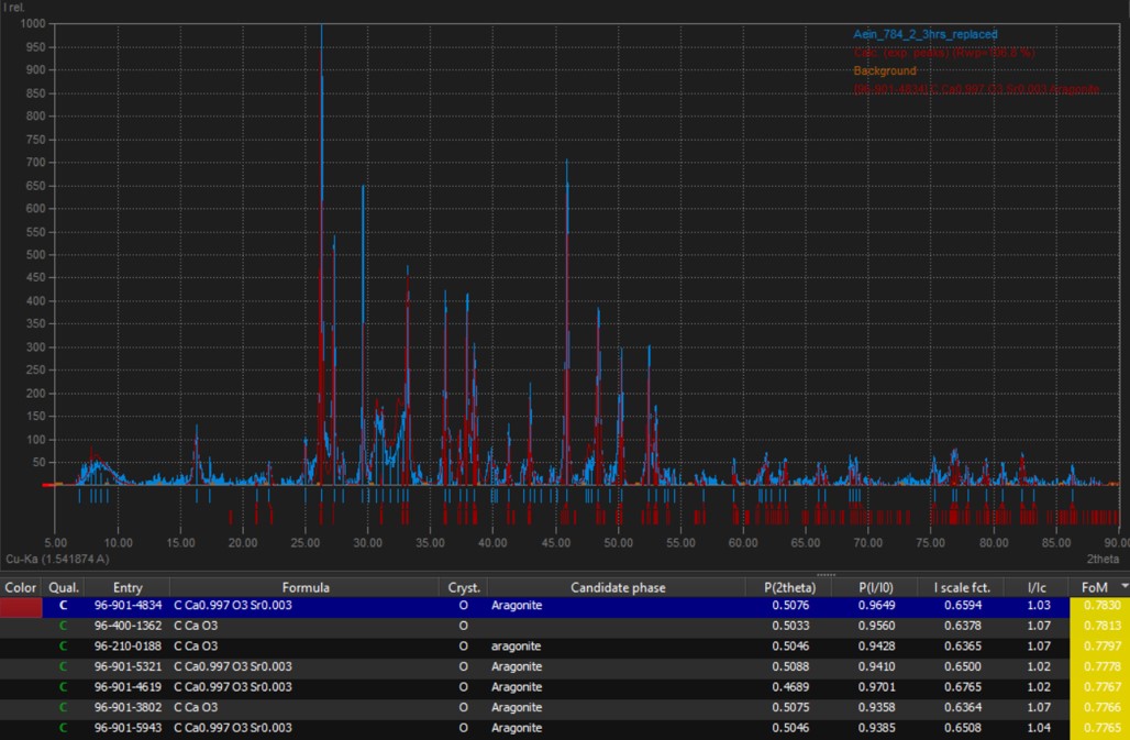
Sample 2, consisting of material obtained from CP1 and CP2 with contamination from CP3 and CP4, shows under PXRD-analysis the presence of aragonite CaCO3 (Fig. 17), a colorless to white or grey mineral, often stained with various hues by impurities, such as blue, green, or red, but colourless in transmitted light.
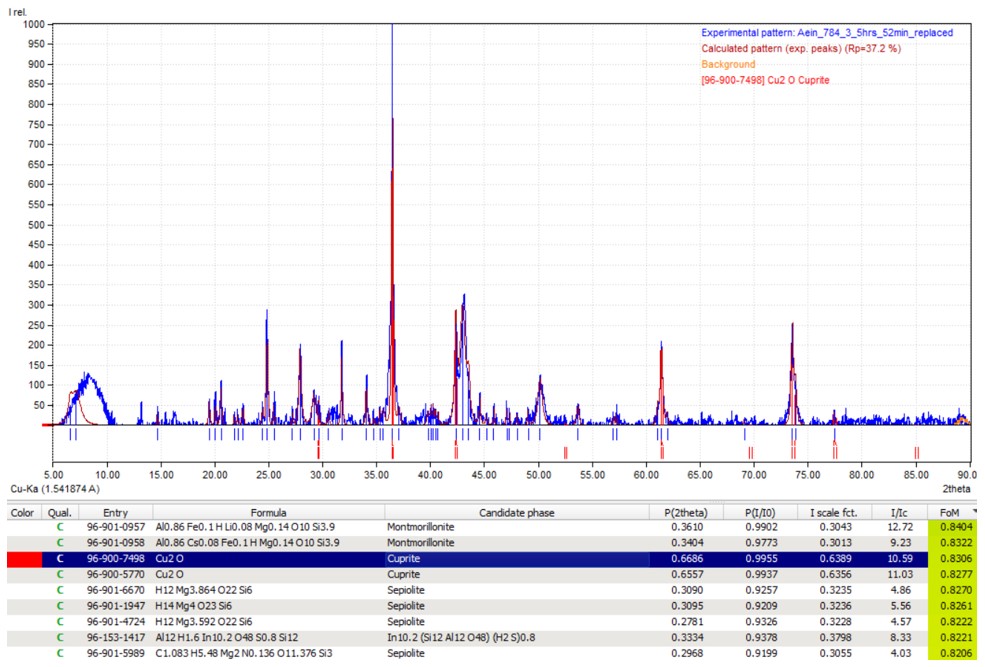
Sample 3, consisting of material obtained from CP3, shows under PXRD-analysis the presence of cuprite Cu2O (Fig. 18), which is consistent with the characteristic red colour under polarized light (Fig. 15). The sample also contains clay minerals montmorillonite and sepiolite.
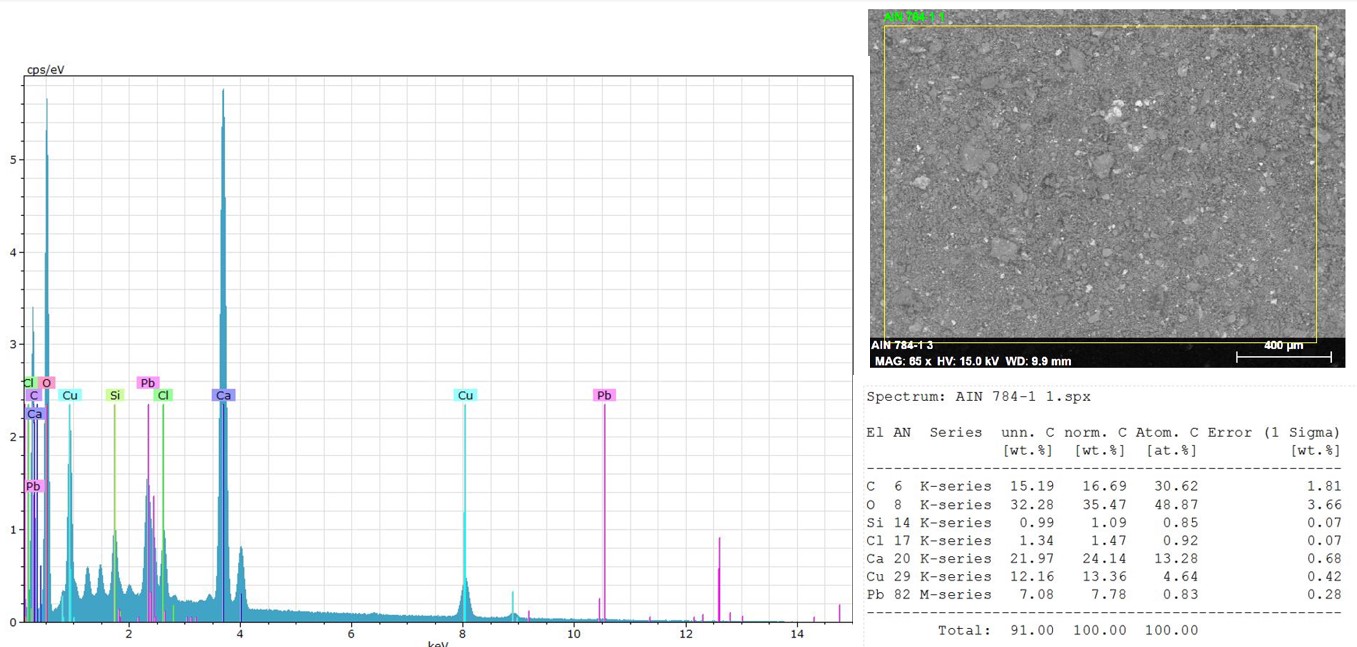
Sample 1 (D1) shows under EDS-analysis a significant amount of calcium with 24,14 % (weight percent) norm. C and some lead 7,78 % norm. C (Fig. 19). This could indicate that the white substance is a precipitation of a calcium compound contaminated with copper ions.
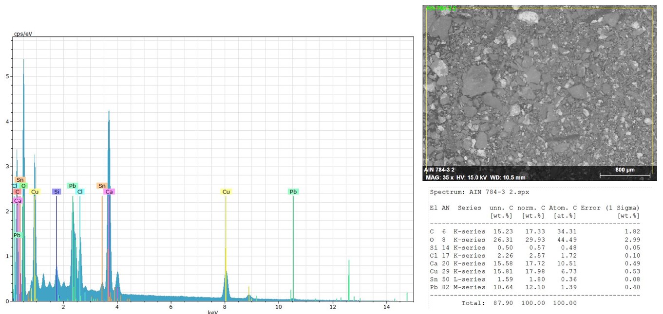
Sample 2 (CP1 and CP2) shows under EDS-analysis somewhat similar values, but a lower content of calcium of 17,72 % (weight percent) norm. C and a higher content of chlorine of 2,57 % norm. C (Fig. 20). This could indicate the presence of copper chloride compounds such as nantokite or copper trihydroxychlorides closer to the metal surface.
 Credit The Royal Danish Academy, I.Langemark.
 Credit The Royal Danish Academy, I.Langemark.
 Credit The Royal Danish Academy, I.Langemark.
 Credit The Royal Danish Academy, I.Langemark.
 Credit The Royal Danish Academy, I.Langemark.
 Credit The Royal Danish Academy, I.Langemark.
 Credit The Royal Danish Academy, I.Langemark.
Multiform
Mostly type I with locally type II (Robbiola)
None.
None.
The figurine is a leaded bronze.
The corrosion products, which cover the entire object, are multi-layered. Most of the corrosion layers correspond to type I of Robbiola.
Three samples were analyzed with PXRD: chlorartinite was detected in a sample obtained from D1. Aragonite was detected in a sample obtained from CP1/CP2, with possible contamination from CP3 and CP4. PXRD-analysis verified CP3 as cuprite mixed with typical clay minerals.
EDS-analysis of material obtained from D1 and CP1 respectively both showed a high content of calcium, some lead, and chlorine. By comparing the data from EDS- and PXRD-analysis, chlorine is presumed to be present in the form of Mg2(CO3)Cl(OH)·2.5H2O, the chloride analog of artinite (Frost et al. 2009), and not as nantokite or the tri-hydroxychloride often associated with active corrosion (Scott 2002).
However, the presence of magnesium compounds such as chlorartinite (Mg2(CO3)Cl(OH)·2.5H2O) and aragonite (CaCO3) may contract water from air humidity, and accelerate corrosion of the metal.
References on object and analytical methods
Reference object
1. Jørgensen, M. (2009) Katalog Ægypten V. Ny Carlsberg Glyptotek 2009, 97.1.
2. MiCorr_Figurine of Egyptian God Bes n°1 - AEIN 223
Reference analytical method
3. Frost, R. L. Bahfenne, S., Graham, J. (2009) Raman spectroscopic study of the magnesium-carbonate minerals-artinite and dypingite. Journal of Raman Spectrocopy, 40 (8). Pp. 855-860. doi: 10.1002/jrs.2152.
4. Scott, D. A. (2002) Copper and Bronze in Art - Corrosion, Colorants, Conservation, Getty Conservation Institute, Getty Publications, 2002.