Pre-Colombian funerary mask VK_D_2256_462
(HE-Arc CR, Neuchâtel, Neuchâtel, Switzerland). (Christian Degrigny, None) & Valentin. Boissonnas (HE-Arc CR, Neuchâtel, Neuchâtel, Switzerland) & Audrey. Pera (HE-ARC CR, Neuchâtel, Neuchâtel, Switzerland)
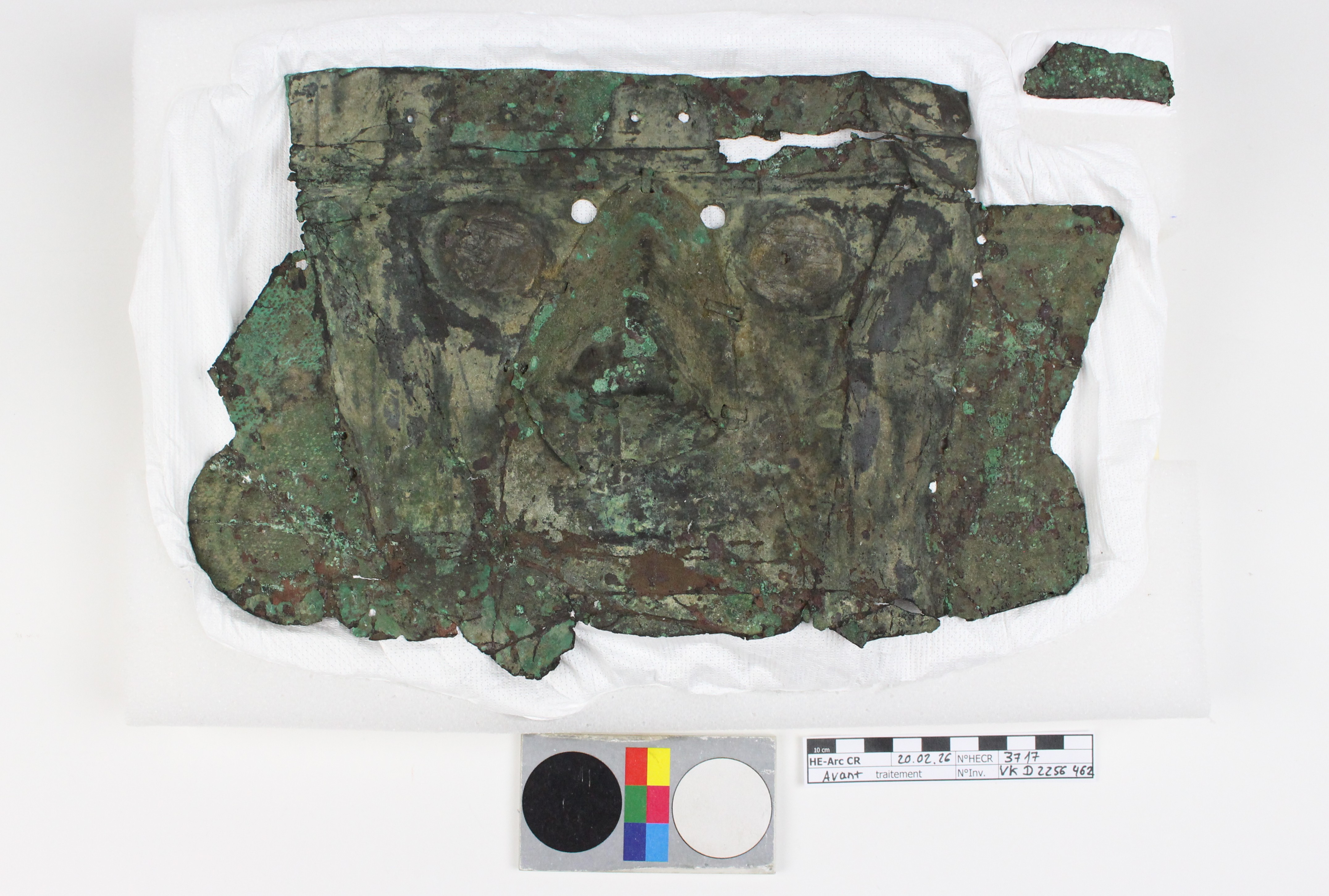
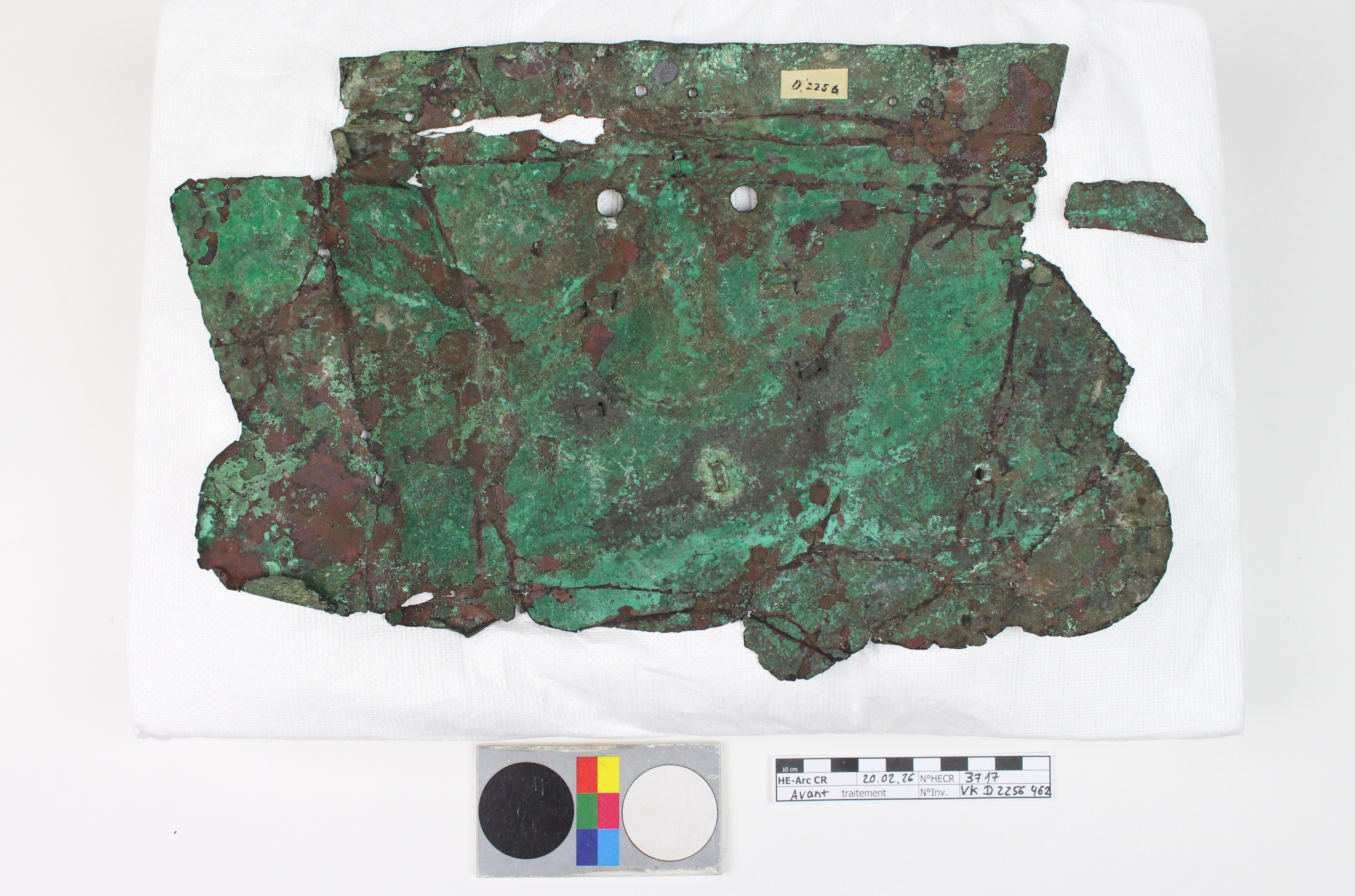
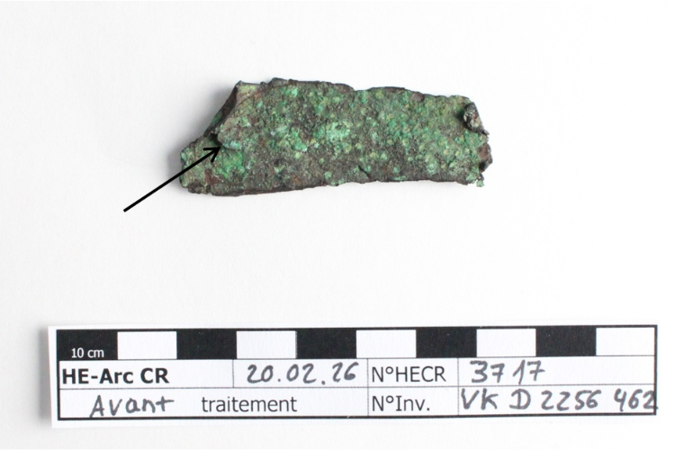
This pre-Columbian funerary mask originates from the Chicama Valley in Peru. It is produced from a single sheet of metal. The nose is a separate piece, attached to the metal sheet by six staples. Blue-green and reddish-brown corrosion products are visible. The burial conditions caused the mask to corrode differently between the sides and the center (Fig. 3). There is a gap of a few centimeters in the upper right section, and a detached fragment—originally from the lower section—is associated with the mask (Fig. 5). Dimensions: Length = 30cm, Width = 19cm, Height = 4cm.
Funeral mask
Chicama valley, Peru
None
Unknown
Unknown
Kulturmuseum St-Gallen
Kulturmuseum St-Gallen
VK_D_2256_462
N/A
By visual observation, we can see some beige fibres, the weave of a textile is printed on the corrosion products and some mineralised fibres are visible. An unknown substance is present on the eyes.
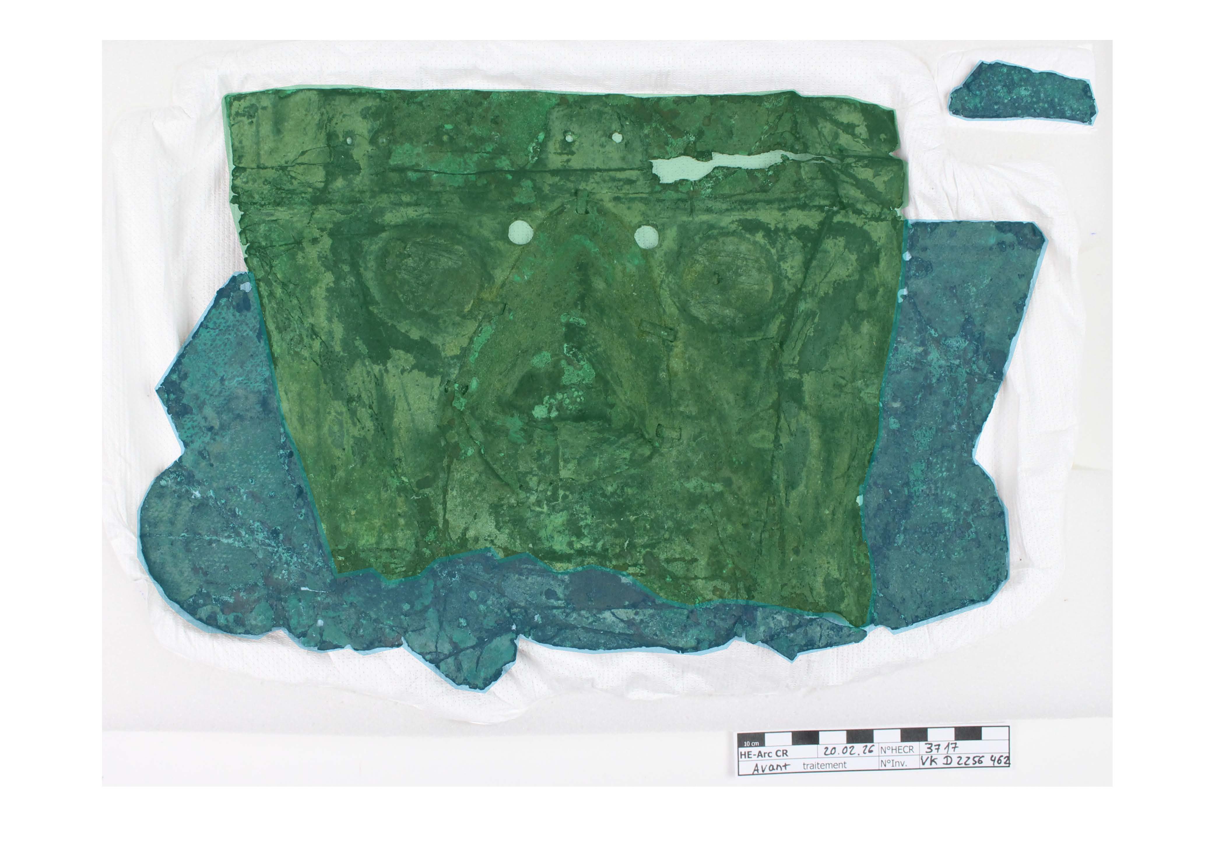 Credit HE-ARC CR, A.Pera.
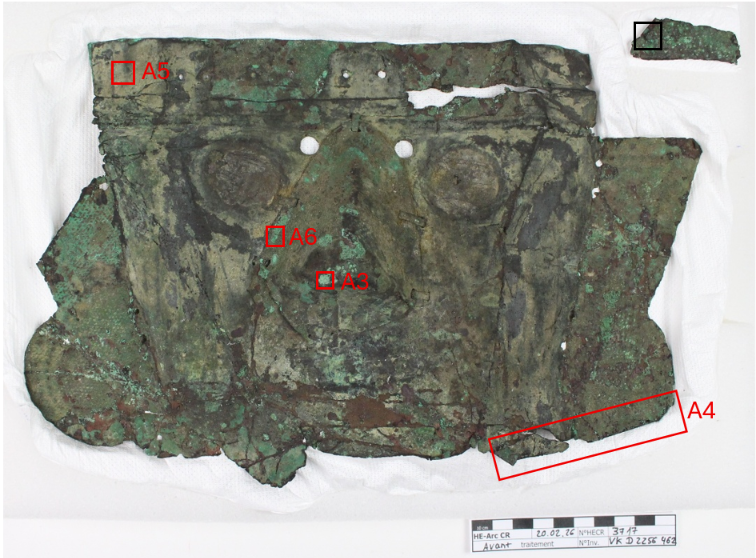 Credit HE-Arc CR, A.Pera.
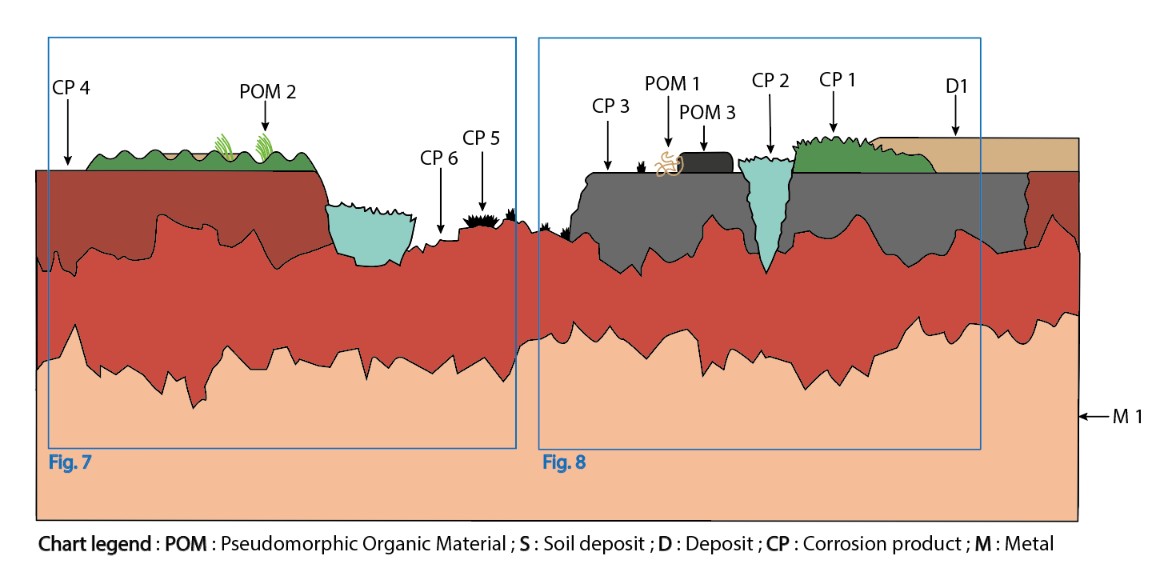
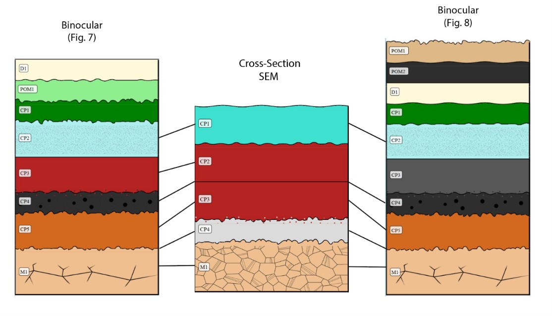
The schematic representation below gives an overview of the corrosion structures encountered on the funeral mask from a first visual macroscopic observation.
| Strata | Type of strata | Principals characteristics |
| D1 | Deposit | Layer, light brown, thin, discontinuous, matte, opaque, friable, soft |
| POM 1 | Organic remains | Light brown, medium, isolated, non-observable brightness |
| POM2 | Organic remains | Light green, thin, discontinuous, matte, opaque, brittle, soft |
| POM3 | Organic remains | Layer, extra dark grey, medium, isolated, matte, opaque |
| CP1 | Corrosion product | Layer, dark green, medium, discountinuous, matte, opaque, compact, severable, hard |
| CP2 | Corrosion product | Lens-shaped, pale turquoise, medium, isolated, matte, opaque, powdery, very soft |
| CP3 | Corrosion product |
Layer, dark grey, thin, continuous, metallic, opaque, compact, malleable, soft |
| CP4 | Corrosion product | Layer, dark red, thin, continuous, matte, opaque, friable, hard |
| CP5 | Corrosion product |
Veins or seams, black, thin, scattered, matt, opaque, non-compact, brittle, soft |
| CP6 | Corrosion product | Layer, dark orange, thin, scattered, matte, opaque, compact, severable |
| M1 | Metal | Layer, light orange, medium, continuous, metallic, opaque, compact, tough, hard |
Table 1 : Description of the characteristics of the strata as observed under binocular.
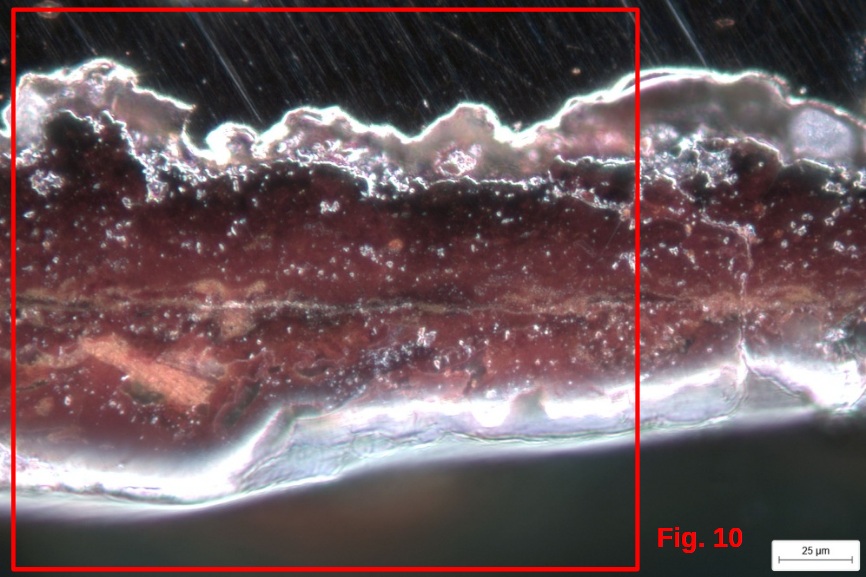 Credit HEI-Arc, S.Ramseyer.
As illustrated on Figs. 4 and 5, the sample was taken on the detached fragment of the mask. The fragment shows a higher degree of mineralization than the rest of the mask (Fig. 11). It may have become detached during burial and been subjected to different burial conditions from the mask. Dimensions: 3x3mm.
Cu As Alloy
Annealed after cold working
None
None
Sampling aimed to study the composition, corrosion, and microstructural properties of the artefact.
It was temporarily embedded in a methyl methacrylate-based resin to examine its cross-section. After the resin was removed, the sample was reintegrated into the object.
Analysis performed :
Non-invasive approach
- XRF, handheld device Thermo ScientificTM Nixton XL3t Goldd +, general metal mode (20s Main / 20s Low / 20 s Light) and mining mode (20s Main / 20s Lw / 20s High / 20s Light)
- X-radiography (59kV, 5 mA, 60s, d = 100cm)
Invasive approach
- SEM-EDS performed on individual samples and on the cross-section using a SEM-FEG JEOL 7001-F equipped with a silicon-drift EDS Oxford detector (Aztec analysis software) with an accelerating voltage of 20kV
- Metallography: the cross-section was observed before and after etching with aqueous chloride iron reagent
- FTIR spectroscopy using Thermo ScientificTM Nicolet iN10MX spectrometer and Thermo ScientificTM OMNIC software
XRF performed directly on the metal surface of the mask with mining mode indicated the presence of arsenic (around 1% in mass).
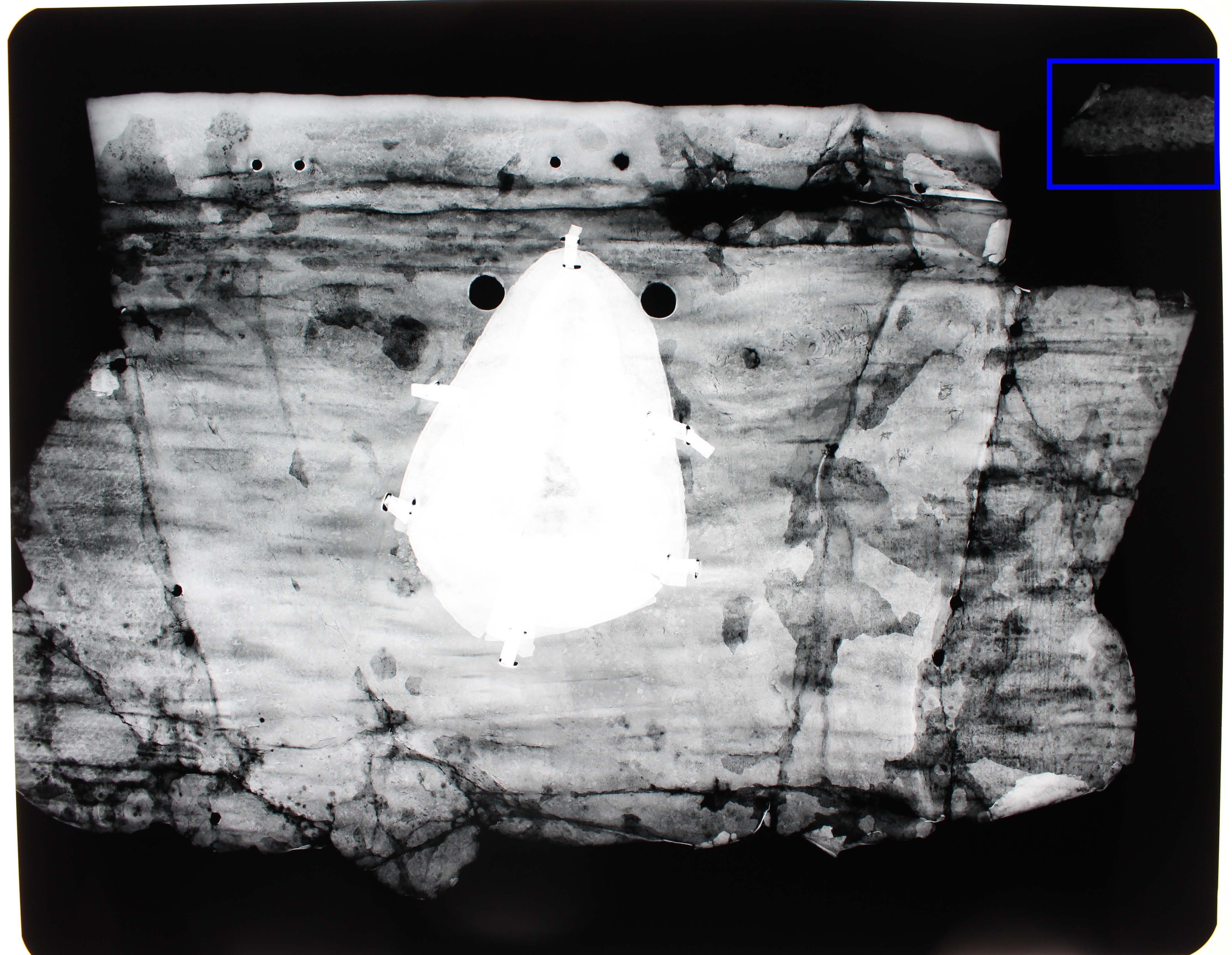
X-ray radiography (Fig. 11) shows traces of hammering and embossed decorations on the ears. Gaps (top right) and areas of reduced density (top left and centre-right) appear darker. The metal at the nose appears denser, but this is due to the overlapping of sheets in that area. Because of its almost entirely mineralisation, the sample is barely discernible (blue rectangle).
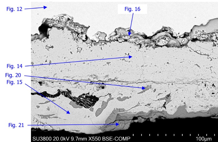
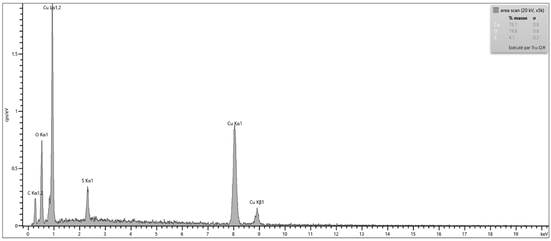
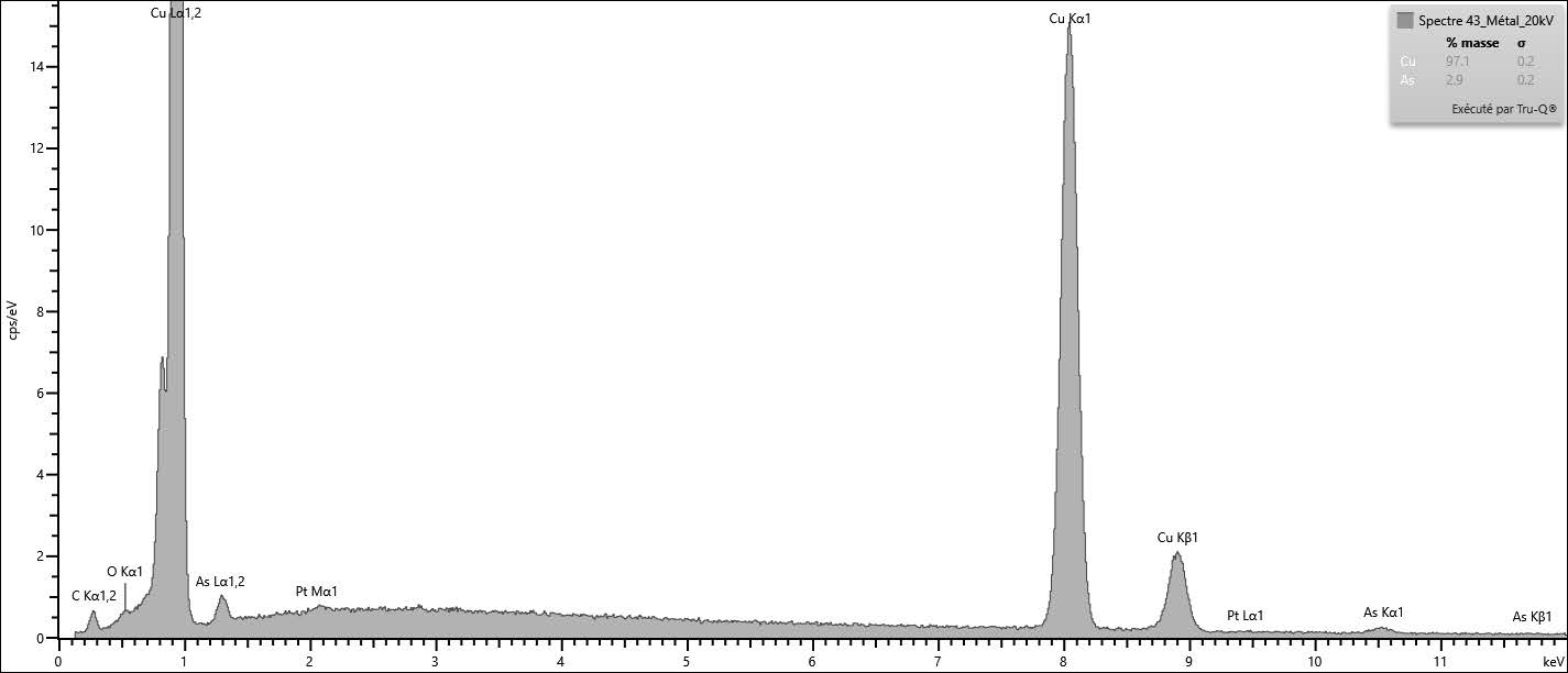
The metal analysed in cross-section with EDS consists of a copper-arsenic alloy (Fig. 12 and Table 2). It confirms the preliminary result obtained with XRF.
| Elements | Cu | As |
| Mass % | 97.1 | 2.9 |
Table 2: EDS analysis of the metal of the mask.
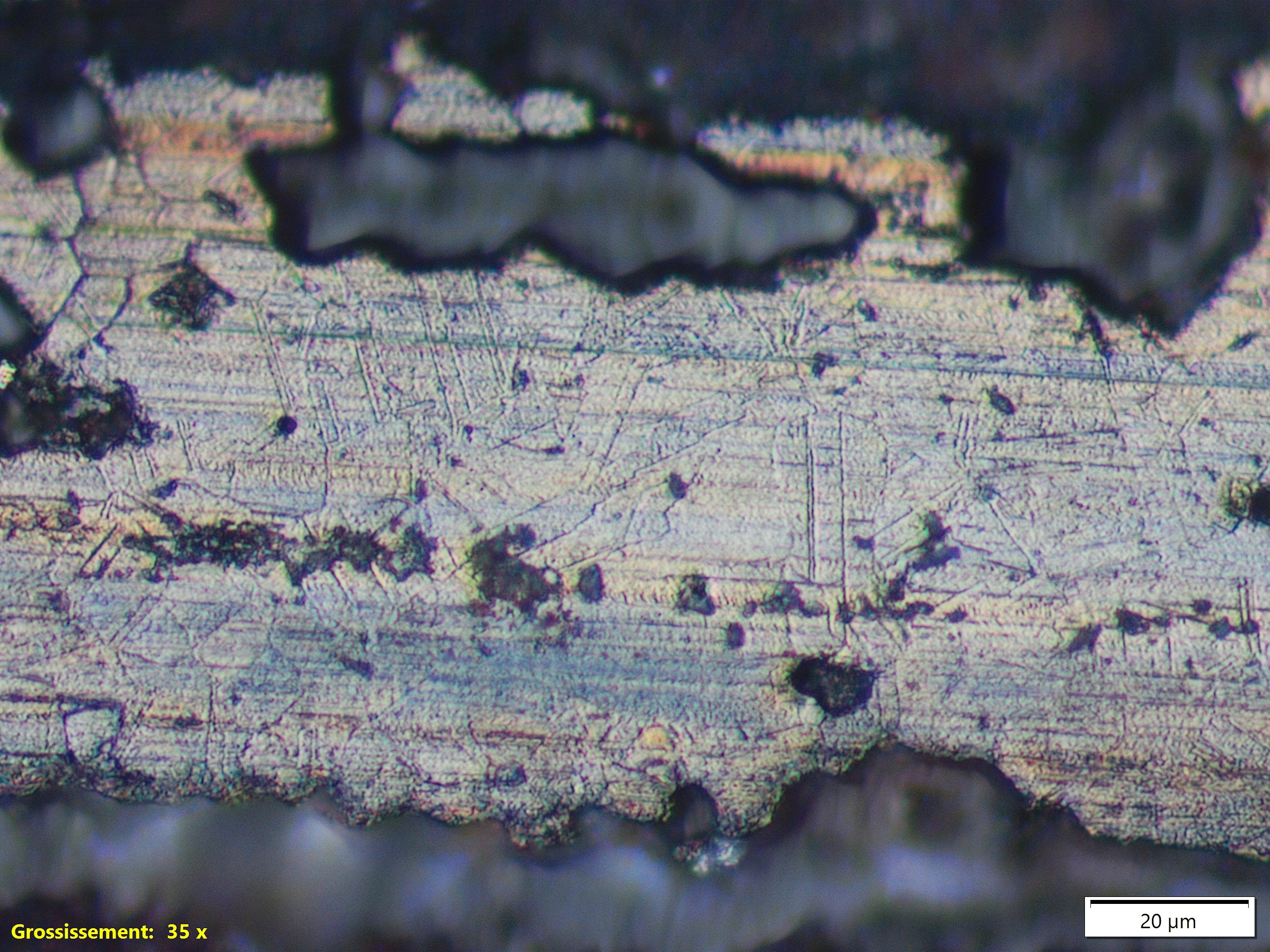
The microstructure of the metal shows some hexagonals grains with twinned lines which indicate that the metal sheet was annealed after cold-working (Scott, 1991) (Fig. 13).
 Credit HEI-Arc, S.Ramseyer.
Large polygonal grains with few twins
Cu
As
None.
The central part of the mask (Fig. 3) features a smooth, grey surface layer (CP3, Fig. 6). In places, across the entire mask, a smooth dark red layer (CP4, Fig 6) can be observed. On the nose and on the upper part of the mask a thick layer of green corrosion product (not analysed) is visible (CP1, Fig. 6). The sides of the mask (around the ears (Fig.3)) present a more irregular surface containing mineralized fibers (POM1, Fig. 6) on the same green corrosion layer (CP1, Fig. 6). Underneath a dark orange layer is present (CP6, Fig. 6) with black, powdery veins (CP5). Pale, powdery blue spots appear scattered (CP2).
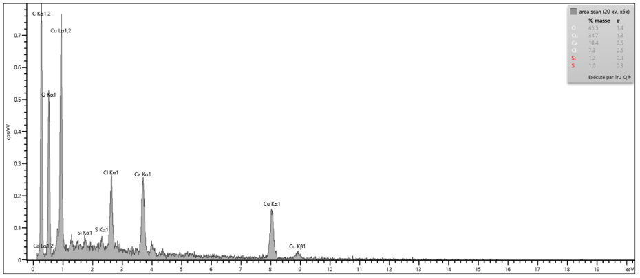
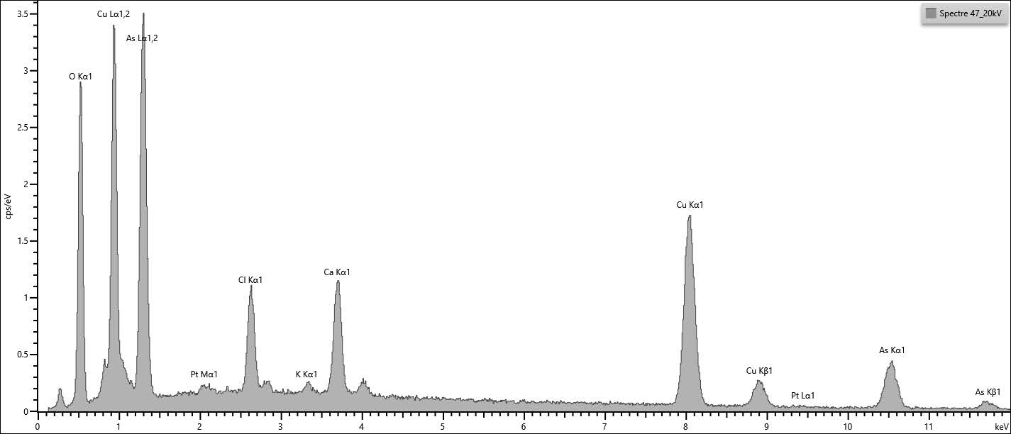
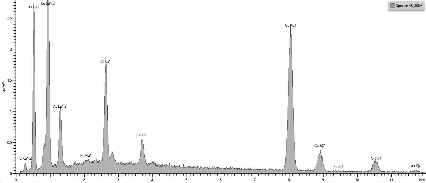
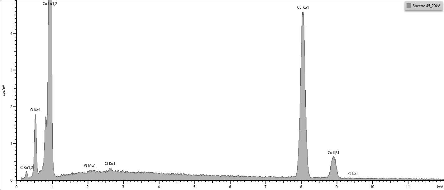
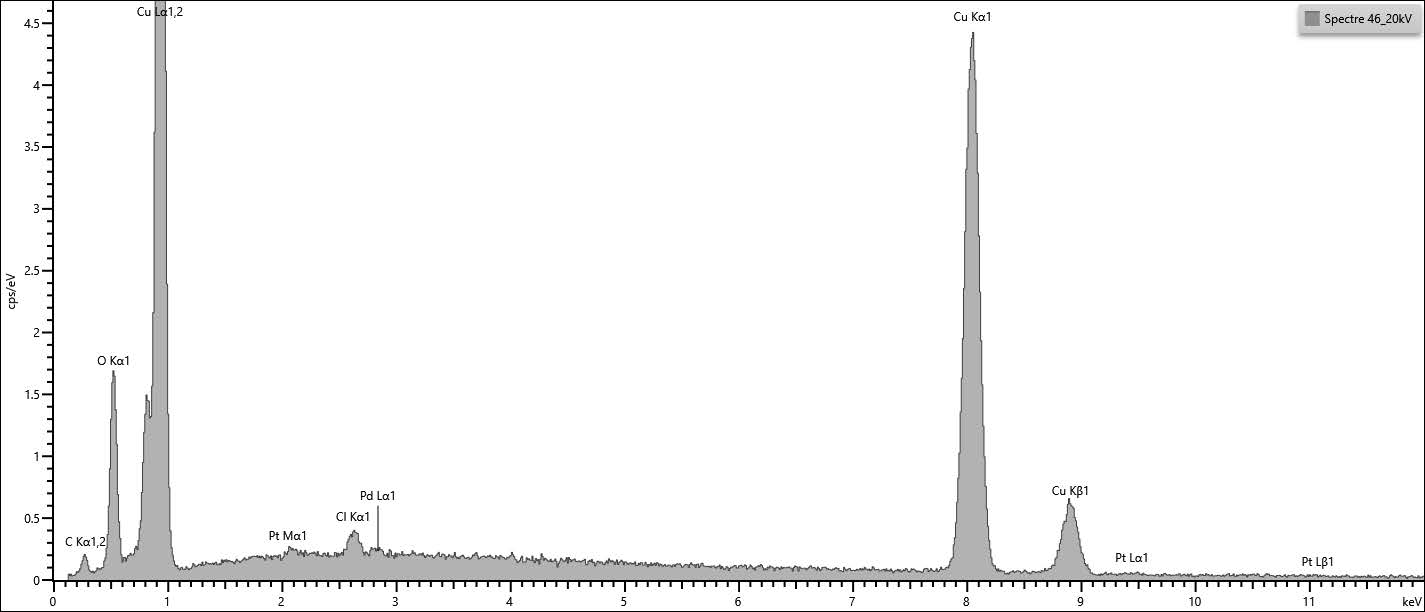
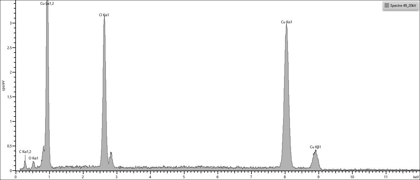
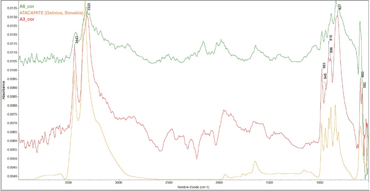
The dark reddish-orange/brown layer (CP4 and CP6, Fig. 6), composed primarily of copper and oxygen, can be identified as cuprite (Figs. 14 and 15). EDS analysis performed at the interface between the remaining metal and the corrosion products reveals the presence of high concentration of chlorides (Fig. 16, nantokite?). FTIR analysis of two areas of powdery, light-blue corrosion products (CP2, Fig. 6) yielded results consistent with atacamite (Fig. 17), a copper hydroxychloride resulting from an active corrosion process.
The object was likely exposed to a sulfur-rich environment, causing the appearance of black spots (Fig. 18). Other elements (Ca and Cl) have been identified on the top surface (Figs. 19 to 21).
 Credit HEI-Arc, S.Ramseyer.
 Credit HEI-Arc, S.Ramseyer.
 Credit HEI-Arc, S.Ramseyer.
 Credit HE-Arc CR, E.Joseph.
Multiform
Mostly type I with locally type II (Robbiola)
None.
The stratigraphies obtained via observation under a binocular microscope and through cross-sectional analysis exhibit several differences. The sample examined in cross-section comes from a detached fragment—originally from the lower part of the mask. It appears to align with the observations made under the binocular microscope, revealing a smooth, dark-red surface layer (Fig. 7), while also displaying a specific CP4 that is rich in chlorides.
In cross-section, CP1, CP2, and CP5 appear similar to CP2, CP3, and CP5 as observed under a binocular microscope. D1, POM1–3, and CP4 do not appear in cross-section, either due to poor adhesion or because they are absent from the sampled area.
The mask is made of a copper-arsenic alloy. The metal sheet underwent annealing after work-hardening. A difference in corrosion products is observed between the edges of the mask and its central section: the center displays a smooth, grey layer and locally, across the entire surface, a smooth dark red layer whereas the edges reveal the imprint of a textile weave as well as mineralized fibers.
The reddish-orange/brown corrosion product was identified as cuprite. The presence of chlorides at the metal/corrosion-product interface prompted further investigation and an analysis of the light-blue, powdery spots found on the surface. These were identified as a hydroxychloride (such as atacamite). Consequently, a process of active corrosion may be underway.
References on object and analytical methods
Scott, D. 1991. Metallography and Microstructure of Ancient and Historic Metals. Marina del Rey, CA : Getty Conservation Institute in association with Archetype Books.