Fragments of oenochoe GV132-01 US26-obj.10
Christian. Degrigny (HE-Arc CR, Neuchâtel, Neuchâtel, Switzerland) & S. Gillioz (HE-Arc CR, Neuchâtel, Neuchâtel, Switzerland) & Valentin. Boissonnas (HE-Arc CR, Neuchâtel, Neuchâtel, Switzerland)
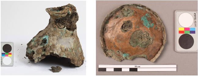
Base of an oenochoe. Dimensions: L = 82 mm; W = 74 (after degradation) ; T =6,5 mm; WT = 30,9 g.
Oenochoe, vessel
Genève, Geneva, Switzerland
Place Simon-Goulart GE, 2012
Roman Times
20 BC _ 50 AC
Soil
Service cantonal d’archéologie, Genève, Geneva
Service cantonal d’archéologie, Genève, Geneva
GV132-01/US26-obj.10
Not conserved
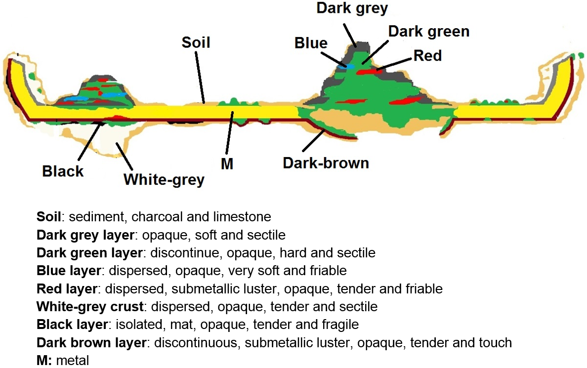
The schematic representation below gives an overview of the corrosion layers encountered on the oenochoe base from visual macroscopic observation.
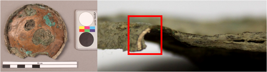
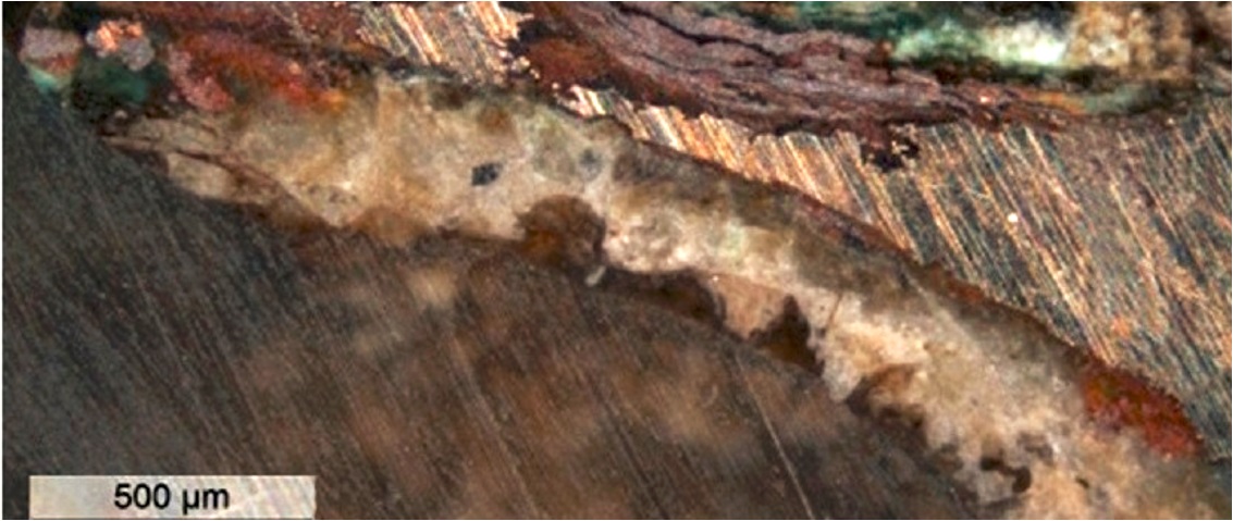
The sample was cut from the edge shown in Fig. 2. Its dimensions are L = 6.5 mm, W = 1.5 mm. The sample contains some remaining metal covered with a crust and a voluminous pustule corrosion (Fig. 4).
Tin Bronze
Cold worked with repeated annealing and final cold working
HECR 1455 – S2
HE-Arc CR, Neuchâtel, Neuchâtel
Musée cantonal d’archéologie et d’histoire, Lausanne, Vaud
2013, metallography and chemical analyses
Analyses performed:
Metallography (etched with ferric chloride reagent), SEM-EDS.
The remaining metal is a tin bronze (Table 1). The etched metal shows a structure of polygonal grains with twin and strain lines (Fig.6).
| Elements | Cu | Sn |
|---|---|---|
| mass% | 91 | 9 |
Table 1: Chemical composition of the metal. Method of analysis: SEM-EDX, Lab of Electronic Microscopy and Microanalysis, IMA (Néode), HEI Arc.
 Credit HEI Arc, S.Ramseyer.
Polygonal grain with twin and strain lines
Cu
Sn
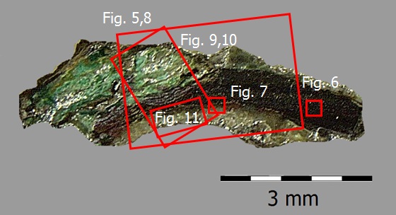
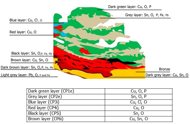
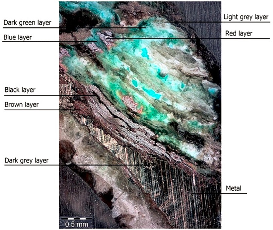
Intergranular corrosion is observed on the edges of the remaining metal (Fig. 7).The sample shows two forms of corrosion: multi-layered pustule corrosion at the left extremity of the sample (Fig. 8, area 1) and a corrosion crust covering the metal (Fig.8, area 2). The multi-layered pustule corrosion has an average thickness of about 1.1 mm (L) and 0.79 mm (W) (Fig.9). It is composed of a sandwich of 7 corrosion products, mainly green, grey, red and blue in dark field. Microscopic observation allows us to highlight new corrosion products that were not detected during the first visual examination (Fig. 9):
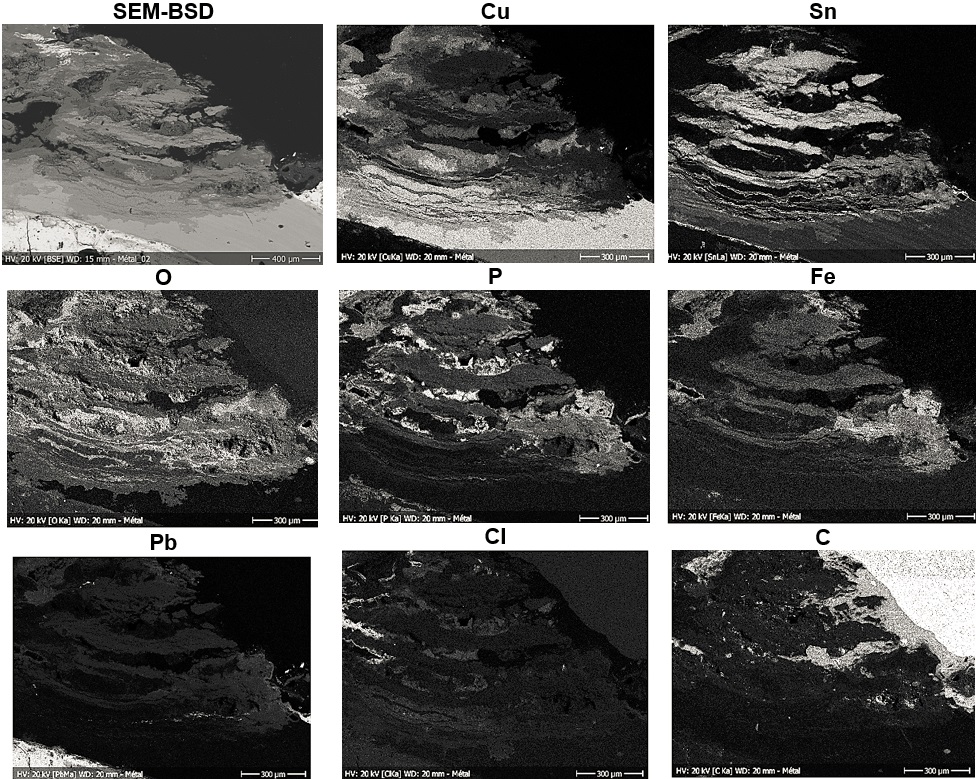
1. Light grey layer, containing mainly Sn, O, some Fe, P and a small amount of Pb (Fig. 10)
2. Dark green layer, containing mainly Cu, O and P (Fig. 10)
3. Red layer, containing mainly Cu and O (Fig. 10)
4. Blue layer, containing mainly Cu, Cl (Fig. 10)
5. Black layer containing mainly Sn, O (Fig. 10)
6. Brown layer containing mainly Cu, Sn, O (Fig. 10)
7. Dark grey layer containing mainly Cu, Sn, O (Fig. 10)
Superior markers such as contextual Si, Fe and P are present in several layers. Their penetration illustrates the cracking of the primary corrosion layer during the formation of the pustule. The P-enrichment in some corrosion layers may be due to an environment rich in organic material (for example bones). The multi-layered pustule corrosion type has developed similarly to the process presented by Formigli (1975, p.53) and Scott (2002, p.337).
The corrosion crust on the metal has an average thickness of about 70 μm (Fig. 11). It consists of two sub-layers. The inner corrosion layer is thin and brown in dark field or light grey in bright field. It has penetrated into the metal structure in some areas (Fig.7). In dark field, the outer corrosion layer is constituted of a heterogeneous light grey corrosion crust (Fig.11). The inner brown corrosion layer is enriched in Sn and O but also contains P, while the outer light grey corrosion layer is mainly composed of Pb and O but also contains P and Fe (Fig. 10). The outer light grey layer is probably due to the presence of a soft solder used to assemble the base to the body.
|
Elements |
Cu | Sn | O | P | Fe | Pb | Cl |
|---|---|---|---|---|---|---|---|
| Grey layer | nd | +++ | ++ | ++ | ++ | + | nd |
| Dark green layer | ++ | nd | +++ | +++ | + | + | + |
| Red layer | +++ | nd | ++ | nd | + | nd | nd |
| Blue layer | +++ | nd | + | nd | nd | nd | +++ |
| Black layer | nd | +++ | +++ | + | + | + | nd |
| Brown layer | ++ | ++ | ++ | nd | nd | nd | nd |
| Dark grey layer | ++ | ++ | ++ | nd | nd | nd | nd |
| Dark brown layer | nd | +++ | ++ | + | + | + | nd |
| Light grey layer | nd | nd | ++ | + | + | +++ | nd |
Table 2: Chemical composition of the multi-layered pustule corrosion from Figs. 9, 10 and 11. SEM-EDX, Lab of Electronic Microscopy and Microanalysis, IMA (Néode) (+++: high concentration, ++ medium concentration, + low concentration, nd: not-detected).
 Credit HE-Arc CR.
 Credit HE-Arc CR.
 Credit HEI Arc, S.Ramseyer.
 Credit HE-Arc CR.
Multiform (warty - uniform) - pitting
Both Formigli (pustules) and type I (Robbiola) otherwise
Based on the analyses carried out, the schematic representation of the stratigraphy of the multi-layered pustule form has been corrected.
The metal of the oenochoe’s base is a tin bronze. The polygonal and twinned grains with strain lines show that the base has been repeatedly cold worked and annealed with a final cold work. The metal is either well preserved or heavily corroded with the formation of pustules that go through the whole thickness of the metal. The limit of the original surface corresponds to the top surface of the dark brown layer. In the presence of a pustule it is highly deformed but discernible by the tin enriched surface. The corrosion is multiform. The well preserved and only lightly corroded areas are of Robbiola type 1 (Robbiola et al. 1998), the pustules however are of the Formigli type (Formigli 1975).
|
References on object and sample |
|
Reference object 1. Gillioz S. (2012) Oenochoé GV132-01/US.26-obj.10, Genève, Place Simon-Goulart, rapport d’intervention. Haute Ecole ARC, Neuchâtel, 2013, non-publié.
Reference sample 2. Gillioz S. (2012) Oenochoé GV132-01/US.26-obj.10, Genève, Place Simon-Goulart, rapport d’intervention. Haute Ecole ARC, Neuchâtel, 2013, non-publié. |
|
References on analytic methods and interpretation |
| 3. Formigli, E. « Die Bildung von Schichtpocken auf antiken Bronzen ». Arbeitsblätter, Heft 1, 1975, p.51 à 74. 4. Robbiola, L., Blengino, J-M., Fiaud, C. (1998) Morphology and mechanisms of formation of natural patinas on archaeological Cu-Sn alloys, Corrosion Science, 40, 12, 2083-2111. 5. Scott, D. A. Copper and bronze in Art, corrosion, colorants, conservation. Getty publications, Los Angeles, 2002. |