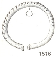
Round bracelet with uniform inclined indentations B3482
Marianne. Senn (EMPA, Dübendorf, Zurich, Switzerland) & Christian. Degrigny (HE-Arc CR, Neuchâtel, Neuchâtel, Switzerland)
Bracelet with uniform inclined rib after Paszthory (1985, 207). Dimensions: Ø = 4cm; WT = 18g (Fig. 1).
Jewellery
Les Eaux-Vives, Genève, Geneva, Switzerland
None
Late Bronze Age
Hallstatt B2/3 (1000BC _ not defined)
Lake
Musées d'art et d'histoire, Genève, Geneva
Musées d'art et d'histoire, Genève, Geneva
B3482
Not conserved
Nothing to report.
Stratigraphic representation: none.
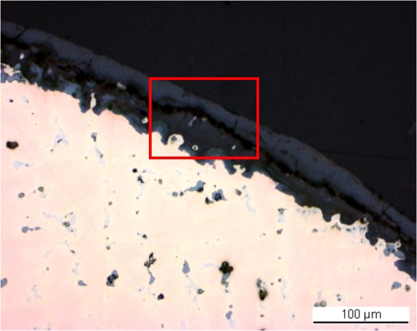
The sample is a section from the central part of the bracelet (Fig. 2). Its dimensions are: L = 2.5mm and W = 0.65mm. The corrosion layer is relatively thin (Fig. 3).
Leaded Bronze
As-cast
MAH 77-110-5
Musées d'art et d'histoire, Genève, Geneva
Musées d'art et d'histoire, Genève, Geneva
1977 and 1991, study of the corrosion layer, metal composition
Nothing to report.
Analyses performed:
Metallography (etched with ferric chloride reagent), Vickers hardness testing, ICP-OES, SEM/EDS.
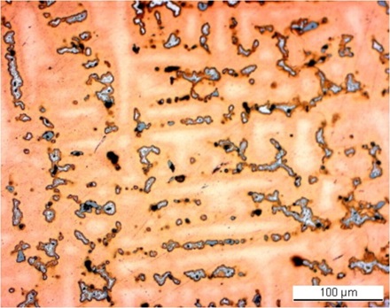
The remaining metal is a porous leaded bronze (Fig. 5 and Table 1). In bright field dark-blue copper sulphide (Fig. 5, Table 2) and tiny dark-grey Pb inclusions (Fig. 5) can be seen. The Sn-rich eutectoid alpha + delta phase appears in light-blue (Fig. 5) and incorporates Pb-rich inclusions. The etched leaded bronze has the dendritic structure of an as-cast metal (Fig. 6) with an average hardness HV1 90. After etching the inclusions have turned darker (Fig. 6) while the eutectoid phase appears whiter.
| Elements | Cu | Sn | Pb | Sb | Ag | Ni | As | Co | Zn | Fe | Bi |
|---|---|---|---|---|---|---|---|---|---|---|---|
| mass% | 90.93 | 6.43 | 1.40 | 0.52 | 0.26 | 0.20 | 0.18 | 0.04 | 0.02 | 0.02 | <0.01 |
Table 1: Chemical composition of the metal. Method of analysis: ICP-OES, Laboratory of Analytical Chemistry, Empa.
|
Elements |
S | Cu | Total |
|---|---|---|---|
| Dark-blue inclusion | 21 | 80 | 101 |
Table 2: Chemical composition (mass %) of dark-blue inclusions on Fig. 5. Method of analysis: SEM/EDS, Laboratory of Analytical Chemistry, Empa.
 Credit HE-Arc CR.
Dentritic structure with pores and inclusions
Cu
Sn, Pb
Nothing to report.
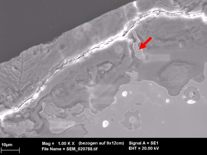
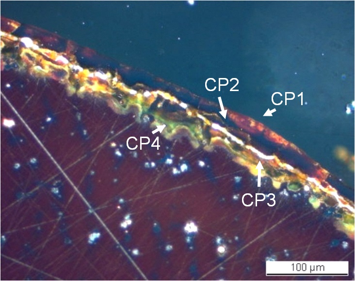
The interface between the metal and corrosion products is irregular (Fig. 5). The corrosion crust has an average thickness of 70µm and is composed of two layers separated by a large fissure (Fig. 5). In bright field, the inner layer includes remnant metal (Sn-rich eutectoid phase, Figs. 5 and 7) and is dark-grey (Fig. 5) while in polarised light it is orange-brownish (CP3, Fig. 8). This Cu depleted layer is rich in Sn, Fe, Si and O (Table 3 and Fig. 8). At the metal - inner layer interface a corrosion product (CP4, light-grey in bright field, greenish in polarised light) shows a slight increase in Cu and Sn content but a decrease of the Fe content (Table 3). In bright field, the outer dense layer is light-grey (CP2, Fig. 5) while in polarised light it appears black with superimposed red to orange areas (CP1, Fig. 8). It is depleted of Cu and richer in Fe. The Sn content is variable but increases in the top brown areas (Table 3 and Fig. 9).
|
Elements |
O | Cu | Sn | Pb | Fe | Si | S | Total |
|---|---|---|---|---|---|---|---|---|
| CP1, outer brown area | 36 | 6.7 | 24 | 2.5 | 27 | 4.3 | < | 102 |
| CP2, outer black layer (average of 2 similar analyses) | 34 | 9.0 | 14 | 2.8 | 34 | 4.3 | < | 99 |
| CP3, inner orange-brown layer (average of 2 similar analyses) | 31 | 16 | 18 | 2.4 | 21 | 5.2 | < | 95 |
| CP4, inner greenish layer (average of 2 similar analyses) | 36 | 29 | 19 | 1.5 | 14 | 6.1 | < | 107 |
Table 3: Chemical composition (mass %) of corrosion layers from Fig. 8. Method of analysis: SEM/EDS, Laboratory of Analytical Chemistry, Empa.
 Credit HE-Arc CR.
 Credit HE-Arc CR.
Fig. 8: Micrograph similar to Fig. 5 and corresponding to the stratigraphy of Fig. 4, polarised light. From bottom left to top right: the metal with blue inclusions and porosities in white, the inner corrosion layer in green, red and orange waves, the fissure in white and the outer corrosion layer in black and red (top zone),
Uniform - selective
Type II (Robbiola)
Nothing to report.
Corrected stratigraphic representation: none.
The leaded bronze shows an as-cast structure. The metal surface is selectively corroded, showing a remnant Sn-rich phase in the inner corrosion layer. Because of this remnant metallic structure, the corrosion type is similar to a type 2 corrosion after Robbiola et al. 1998. In this case, two corrosion processes have occurred in parallel: a typical Cu depletion and Sn enrichment, but at the same time a surface enrichment with Fe and Si that could be explained by an Fe-rich lake environment.
|
References on object and sample |
|
Reference object 1. Paszthory, K. (1985) Der bronzezeitliche Arm- und Beinschmuck in der Schweiz. PrähistorischeBronzefunde X-Bd. 3, München, 243, Tafel 137.Reference sample 2. Empa report 137'695/1991, P. Boll.3. Rapport d'examen, Laboratoire Musées d'art et d'histoire, Genève (1977-110), 1977 and 1991. |
|
References on analytic methods and interpretation |
| 4. Robbiola, L., Blengino, J-M., Fiaud, C. (1998) Morphology and mechanisms of formation of natural patinas on archaeological Cu-Sn alloys, Corrosion Science, 40, 12, 2083-2111. |