Military ID tag 5668-MXX-1008-1
Christian. Degrigny (HE-Arc CR, Neuchâtel, Neuchâtel, Switzerland) & Marie-Jeanne. Scholl (HE-Arc CR, Neuchâtel, Neuchâtel, Switzerland) & Valentin. Boissonnas (HE-Arc CR, Neuchâtel, Neuchâtel, Switzerland)
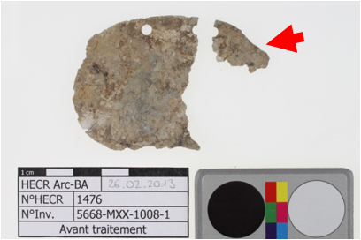
Fragments from an oval shaped German military plate with two circular holes at the central upper edge (Fig. 1). Information concerning the soldier were given in the centre of the plate. The plate is covered with sediments. Dimensions (70 x 50 mm initially): L = 65mm; W = 50mm; T = 1 mm; WT = 6.3g.
Military object
German army, 94th Infantry Regiment, 1918, Carspach, Alsace, France
Recovered in 2011
Modern Times
First World War
Soil
Pôle d’Archéologie Interdépartemental Rhénan, Sélestat, Alsace
Pôle d’Archéologie Interdépartemental Rhénan, Sélestat, Alsace
5668-MXX-1008-1
Not conserved
This 1915 model is the oldest standardized model (Directive 594 of the Ministry of War referenced 1085/7.15.B3). Burial conditions (loess sediments, collapsed shelter (Killianstollen) in March 1918 after a French bombing buried the tag at a depth between 3.5 and 6 meters).
The schematic representation below gives an overview of the corrosion layers encountered on the military tag from a first visual macroscopic observation.
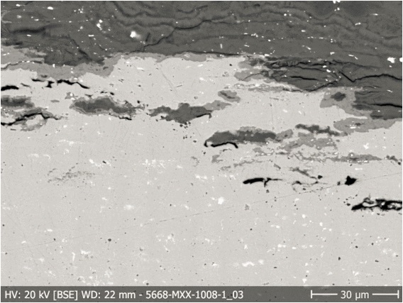
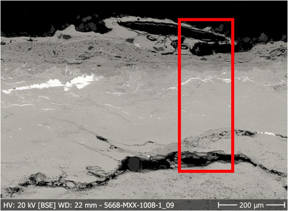
This sample was cut out of fragment b in Fig. 1. The cross-section shows the entire thickness of the plate (L = 8 mm, W= 1 mm). The corrosion goes through the whole metal thickness but a metallic core remains (Fig. 4).
Zn Pb Alloy
Cold worked (laminated)
-
HE-Arc CR, Neuchâtel, Neuchâtel
Pôle d’Archéologie Interdépartemental Rhénan, Sélestat, Alsace
2013, metallography and chemical analyses
Nothing to report.
Analyses performed:
Metallography (etched with HCl 10 M reagent), XRF, SEM/EDS, FTIR and Raman spectroscopy.
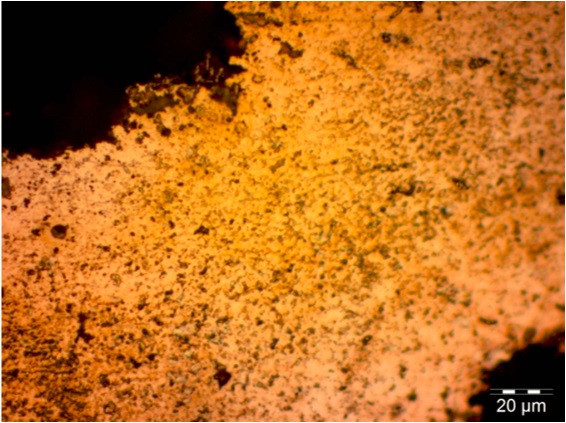
The remaining metal is an almost pure zinc alloy (Table 1) containing 1.8% in weight lead. Lead appears in the metal in the form of small flattened white Pb inclusions (Figs. 7 and 8), confirming the manufacturing technique, namely lamination. The etched metal does not show any specific microstructure (Fig. 9).
| Elements | Zn | Pb |
|---|---|---|
| mass% | 98.2 | 1.8 |
Table 1: Chemical composition of the metal. Method of analysis: SEM-EDS, Lab of Electronic Microscopy and Microanalysis, IMA (Néode), HEI Arc.
 Credit HEI Arc, S.Ramseyer.
?
Zn
Pb
Nothing to report.
The corrosion spreads on the entire thickness of the plate and different forms of corrosion within the same plate may occur (multiform corrosion). Sometimes the metal core still remains, sometimes not.
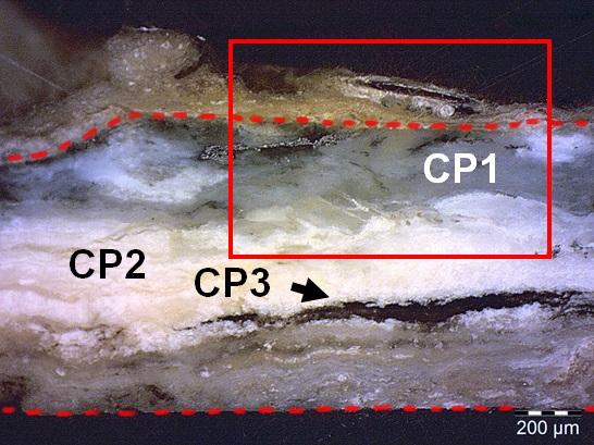
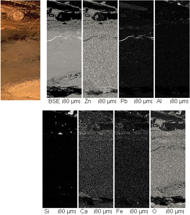
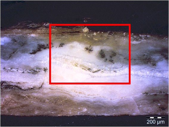
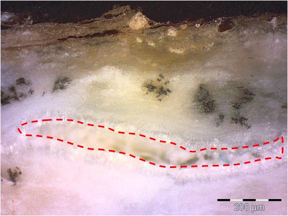
The corrosion structure is multi-layered: several corrosion products overlap. However they are not all continuous layers, since some of them are isolated. Most of the corrosion products layers show structural voids (up to 100 µm thick) and some cracks inside. Three layers can be distinguished (Figs. 5 or 6 - only Fig. 5 is detailed below - and 10) which have the same elements but in different proportions (Figs. 11, 12 and table 2):
- Inner grey layer – CP1 (up to 1 mm thick) is a translucent, compact layer and constitutes most part of the corrosion products. Corroded metal appears inside this layer as lead inclusions within a black corrosion product (Figs. 13 and 14). Sulphur has also been detected by SEM-EDS inside this layer, near the surface, where the colour turns into grey-orange (zinc sulfide or sulphates but to be confirmed, table 2).
- Inner white layer – CP2 (up to 0.6 mm thick) is an opaque and powdery heap, occurring in different places inside inner grey layer and sometimes near the surface, as pitting corrosion. The interface between inner layers grey and white is diffuse. Localized aggregates showing a crystalline microstructure of thickness of 50 µm are located against the walls of a structural void, inside the white corrosion product (Fig. 14).
- Inner black layer – CP3 (up to 0.1 mm) is an opaque and compact layer with a composition similar to inner white layer (Table 2).
On top the metal is covered with non-metallic materials (NMM1, fine sand (10%), silt (75%), clay (15%), vegetal remains) and elements from the corpse (hairs, tissue fibers)). An outer corrosion product has not been identified, but may occur, because zinc combined with sulphur was detected at the surface (Table 2).
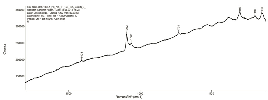
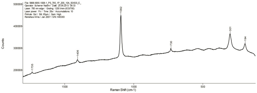
The inner grey layer (CP1, Figs. 5 and 10) has been identified by FTIR as a zinc carbonate (smithsonite), mixed with other unidentified corrosion products. Raman spectra seems to confirm it (Fig. 15). The exact composition of the inner white layer (CP2) is unknown. It shows a bluish fluorescence under UV light and has been identified by FTIR as a mixture of zinc oxides and carbonates, that could be zinc hydroxycarbonates (hydrozincite). Raman spectroscopy shows also a mixture of carbonates and other corrosion products (Fig. 16). The inner black layer (CP3) is a zinc carbonate as well, identified by FTIR and Raman spectroscopy (Fig. 17) appearing as a vein between layers of grey and white (Fig. 10).
|
Elements |
O | Zn | Pb | S |
|---|---|---|---|---|
| Inner grey layer (CP2) | ++ | +++ | + | |
| Black inclusions | + | +++ | ||
| Inner white layer (CP1) | ++ | +++ | + | |
| Inner black layer (CP3) | ++ | +++ | + | |
| Inner grey-orange layer | + | ++ | ++ | |
| Remnant metal phase | +++ | + |
Table 2: Chemical composition of the different corrosion layers from Fig. 13. Method of analysis: SEM-EDS, Lab of Electronic Microscopy and Microanalysis, IMA (Néode) (+++: high concentration, ++ medium concentration, + low concentration, nd: not-detected).
 Credit HE-Arc CR.
Fig. 10: Micrograph of the cross-section, from Fig. 4 (detail) and corresponding to the stratigraphy of Fig. 5, unetched, dark field, 50x. View of the inner layers and the limit of the original surface (red line). We can see that the interfaces between the different layers are not well defined: some of the white corrosion product is found inside the inner grey layer. The micrograph of Fig. 11 is marked by a rectangle,
 Credit HEI Arc, S.Ramseyer.
 Credit HEI Arc, S.Ramseyer.
 Credit HE-Arc CR.
 Credit HE-Arc CR.
 Credit HKB.
Fig. 15: Raman spectrum of the inner grey layer (CP1, ZnCO3 – smithsonite?). Settings: laser wavelength 532nm, acquisition time 10s, one accumulation, filter D2 (0.75-0.8mW), hole 500, slit 80, grating 600. Method of analysis: Raman spectroscopy, Art Technological Laboratory, Departement Conservation HKB,
 Credit HKB.
Fig. 16: Raman spectrum of the inner white layer (CP2, zinc hydroxycarbonate?). Settings: laser wavelength 532nm, acquisition time 10s, one accumulation, filter D2 (0.75-0.8mW), hole 500, slit 80, grating 600. Method of analysis: Raman spectroscopy, Art Technological Laboratory, Departement Conservation HKB,
 Credit HKB.
Multiform - transgranular
?
Nothing to report.
Based on the analyses carried out, the schematic representation of the stratigraphy of the military tag has been corrected.
The military tag is made of a laminated sheet of zinc-lead alloy. The corrosion has entirely penetrated the objet leaving though some spots of remaining original metal inside the corrosion products. Lead particles are not affected by the corrosion process. The main corrosion products are zinc carbonates (smithsonite) and hydroxycarbonates (hydrozincite) but mixtures of different corrosion products can occur. These corrosion products are intermixed and heavily cracked.
|
References on object and sample |
|
Reference object 1. Scholl, M.-J. (2013) Caractérisation des plaques d’identification militaires en zinc provenant du site de Carspach (Alsace, Haut-Rhin, F). Travail de Bachelor Filière Conservation-restauration, Haute Ecole Arc de Conservation-restauration, Neuchâtel.Reference sample 2. Scholl, M.-J. (2013) Caractérisation des plaques d’identification militaires en zinc provenant du site de Carspach (Alsace, Haut-Rhin, F). Travail de Bachelor Filière Conservation-restauration, Haute Ecole Arc de Conservation-restauration, Neuchâtel. |
|
References on analytic methods and interpretation |
| 3. Moser, Z., et al. (1994) The Pb-Zn (Lead-Zinc) System. In Journal of Phase Equilibria, vol.15, n°6, p.643-644. 4. Goodwin, F. E. (2010) “Corrosion of Zinc and its Alloys”. In Cottis, R. A. Shreir’s corrosion. vol.3 Corrosion and degradation of engineering materials. Elsevier Science, Oxford, 2078-2093. 5. De Zoubov, N. et Pourbaix, M. (1963) « Zinc », In Pourbaix, Marcel. Atlas d'équilibres électrochimiques. Gauthier-Villars, Paris, 406-413. |