Roof gutter element
Christian. Degrigny (HE-Arc CR, Neuchâtel, Neuchâtel, Switzerland) & Mathea. Hovind (University of Oslo, Department of archaeology, conservation and history (IAKH-UiO), Oslo, Oslo, Norway)
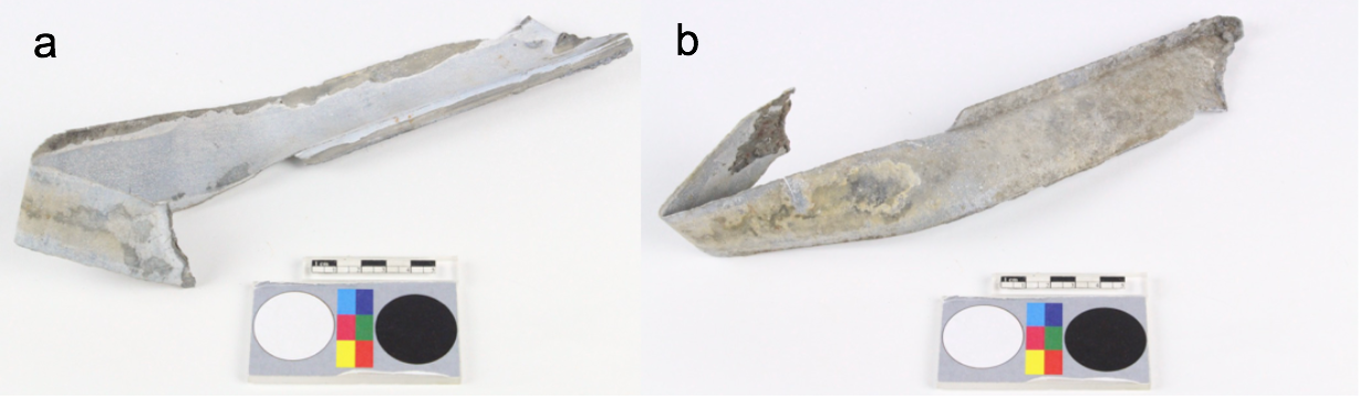
A roof gutter element with remains of lead solder (Fig. 1). Its surfaces are covered by a white, strongly adherent oxide layer. Surface “b” is slightly darker in colour than surface “a” due to the presence of soil particles. Dimensions: L = 265mm; W = 47mm; T = 1mm; WT = 93g.
Architectural element
Château de Germolles, Mellecey, Bourgogne, France
Unknown
Modern Times
19th-20th century
Outdoor atmosphere
Haute Ecole Arc Conservation-Restauration
Château de Germolles, Mellecey, Bourgogne
None
Not conserved
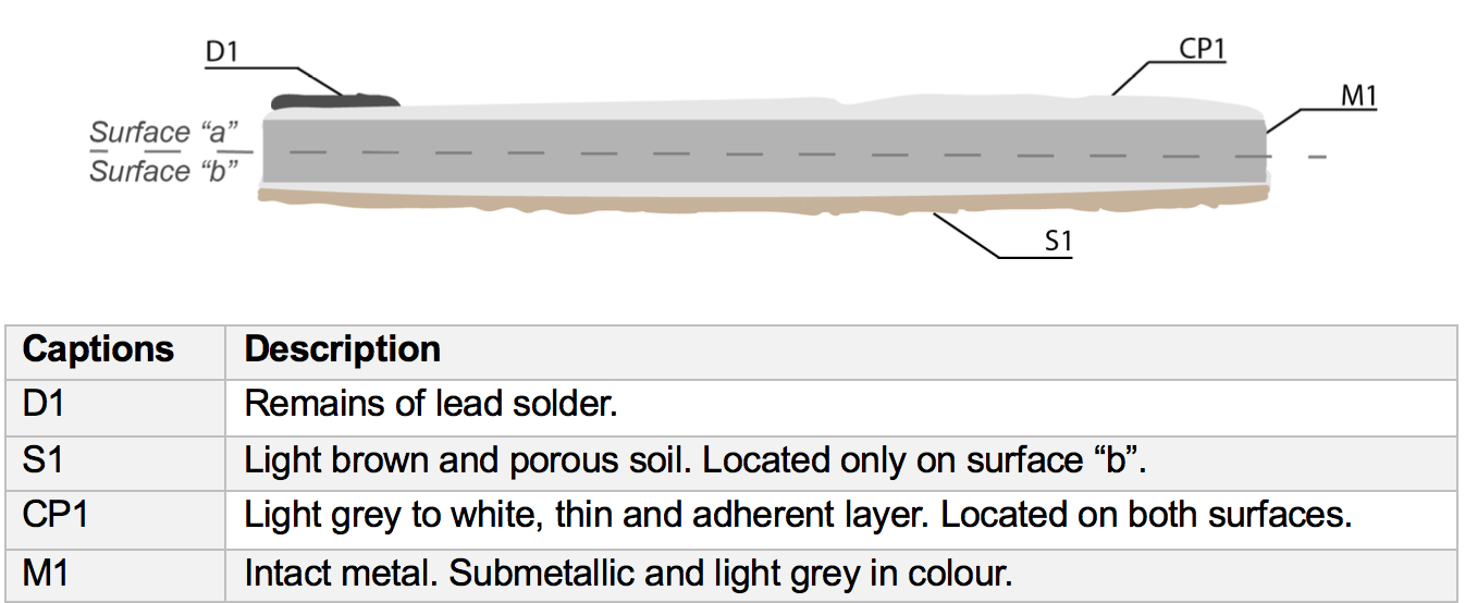
Nothing to report.
The schematic representation below (Fig. 3) gives an overview of the corrosion layers encountered on the object from a first visual macroscopic observation.
A cross-section (Fig. 4) was cut out where the metal is at an angle of approximately 90° to the flat surface. Its upper edge corresponds to surface “a” and contains remains of solder, while the lower edge corresponds to surface “b”.
Zn Alloy
Annealed after (hot) rolling
ZG2018 (zinc gutter sampled in 2018)
Haute Ecole Arc Conservation-Restauration
Haute Ecole Arc Conservation-Restauration
March 2018, metallography and chemical analyses
The fact that the fragment was considered a test material enabled extensive sampling that would not otherwise be possible.
Analyses performed:
Metallography: microscope: Leica DMi8 (a metallographic, inverted, reflected light microscope) with magnification up to 500X. Camera: Olympus SC50 connected to the software “Olympus Stream”, version 1.9.4. Illumination modes: bright field and cross-polarized light. The metal is unetched.
SEM-EDS: instrument: Jeol 6400; voltage: 20 kV; working distance: 18 and 24mm; sample preparation: palladium depot.
XRD: diffractometer system: XPERT-3; Sample stage: Reflection-Transmission Spinner PW3064/60; Anode material: Cu.
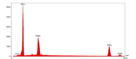
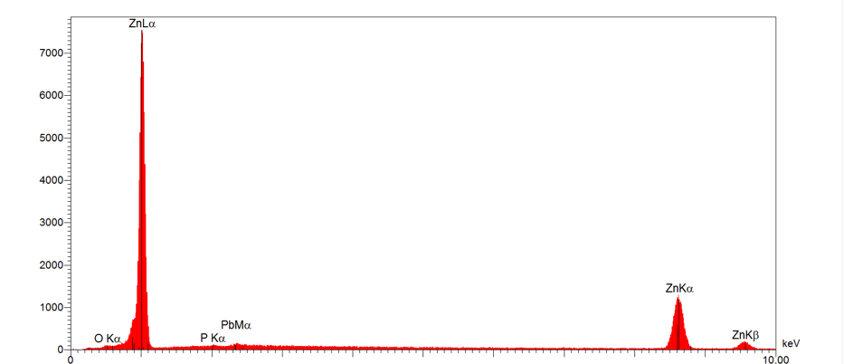
The roof gutter element is composed of Zn with P and Pb at lower concentrations (Fig. 6). The latter is probably originating from the solder that was applied to adjoin several metal sheets for the roof gutter.
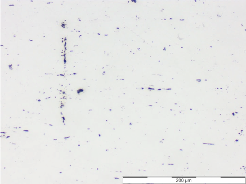
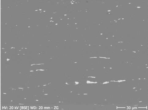
The metal appears white to light grey under bright field (Fig. 7). Under polarized light however (Fig. 8), the microstructure of the metal is visible as small polygonal grains appearing in various shades of brown, a coloured effect due to the anisotropic properties of the metal (Scott 1991: 49). In the SEM-image, white elongated inclusions are visible (Fig. 9) consisting mainly of Zn and Pb with some O (Fig. 10).
 Credit HEI Arc, C.Csefalvay.
 Credit UiO-IAKH, M.Hovind.
 Credit UiO-IAKH, M.Hovind.
 Credit HEI Arc, C.Csefalvay.
Recrystallized structure (polygonal grains)
Zn
P, Pb
Nothing to report.
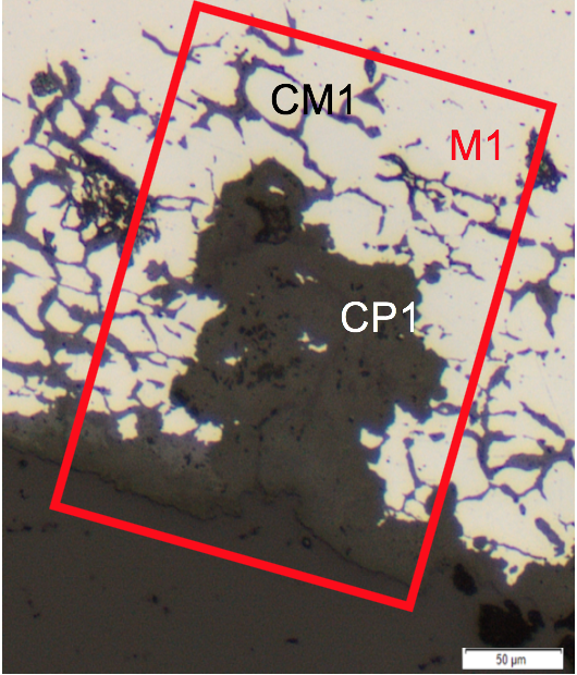
The metal is suffering from intergranular corrosion visible as a white corrosion product (CP1) located within crevices and along the grain boundaries (Figs. 11 and 12). Both the porous and powdery corrosion product (CP1) and the corroded metal (CM1) appear dark grey in bright field (Fig. 11) and white-grey under polarized light (Fig. 12). They both consist of Zn and O (Table 1). The external corrosion product (CP1) contains some S (and not Pb as suggested by Table 1 and Fig. 14) which is probably due to atmospheric pollution. The structural composition of the corrosion product was determined by crystallographic analysis (XRD) to consist of a mixture of zincite (ZnO) and pure zinc (Zn) (Table 2, Fig 13). Mapping of the corroded area by SEM-EDS displays a similar composition but shows additionally the presence of Cl in some of the veins of the corroded metal (Fig. 14). S1 (soil material) is found on the very top of CP1.
|
Elements mass % Layer |
C |
O |
Zn |
Pb |
S |
Al |
Si |
Fe |
Cu |
Sn |
|
CP1, white corrosion product |
7 |
33 |
51 |
3 |
5 |
- |
0.3 |
0.1 |
0.1 |
0.4 |
|
CM1, intergranular corrosion |
6 |
22 |
68 |
3 |
0.1 |
0.1 |
0.1 |
0.1 |
0.1 |
0.5 |
Table 1: Chemical composition of the corrosion layers from Fig. 11. Method of analysis: SEM-EDS. Lab. of Electronic Microscopy and Microanalysis, Néode, HEI Arc, credit MiCorr_HEI Arc, C.Csefalvay. *The sum is the calculated average of three analyses of the same feature, but in different areas.
|
Stratum |
Components* |
|
CP1 |
Zincite (ZnO), Zinc (Zn) |
Table 2: Summary of the results from the crystallographic analysis of the white corrosion product (CP1). A representative spectrum is given in Fig. 13. Method of analysis: XRD. Center of X-ray Analytics, Empa-Swiss Federal Laboratories for Materials Science and Technology (Dübendorf), credit MiCorr_Empa, Z.Balogh-Michels. *The results are based on the analysis of two powder samples from different areas (see Fig. 2 for sample locations).
 Credit UiO-IAKH, M.Hovind.
Fig. 11: Micrograph of the metal sample from Fig. 4 (detail). Unetched, bright field. The corrosion product (CP1) is located at the surface of the metal and in its crevices. Intergranular corrosion (CM1) is visible as thin grey lines in between the metal grains (M1). The area selected for elemental chemical distribution (Fig. 14) is marked by a red rectangle,
 Credit UiO-IAKH, M.Hovind.
 Credit Empa, Z.Balogh-Michels.
Multiform - intergranular
None
Nothing to report.
The schematic representation of corrosion layers integrating additional information based on the analyses carried out is given in Fig. 15.
The roof gutter element consists of Zn with some Pb from the lead solder. It is covered by a strongly adherent layer of zincite (a zinc oxide), indicative of exposure to an unpolluted environment. Still, the metal exhibits intergranular corrosion, a deterioration phenomenon indicative of aggressive conditions. This can possibly be explained by the different locations of the samples. The cross-section studied is likely to correspond to the surface that was exposed to the atmosphere (surface “b”), while the powder samples were from the rear, unexposed surface (surface “a”). Exposure to moisture and low pH in the form of acidic rain are environmental parameters that would encourage intergranular corrosion. Furthermore, the presence of Cl inside the cracks could be due to pollution and its rather concealed location implies that it would be retained inside the metal and not washed away by rain (Selwyn 2004:153-154).
| References on object and sample |
|
References sample |
|
References on analytic methods and interpretation |
|
|