Funerary flask VK_D_1783
Vjosa. Ferati (HE-Arc, Neuchâtel, Neuchâtel, Switzerland)
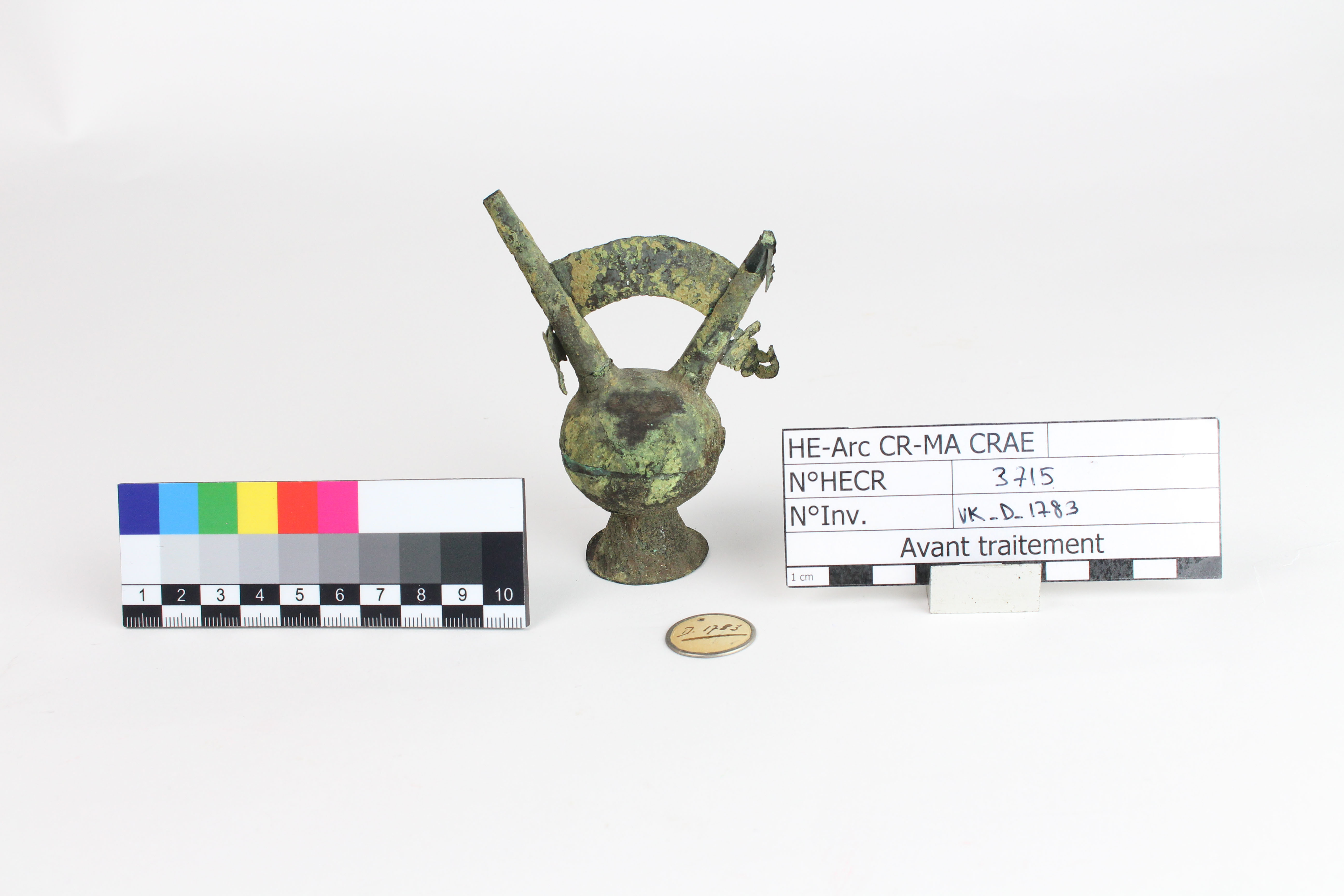
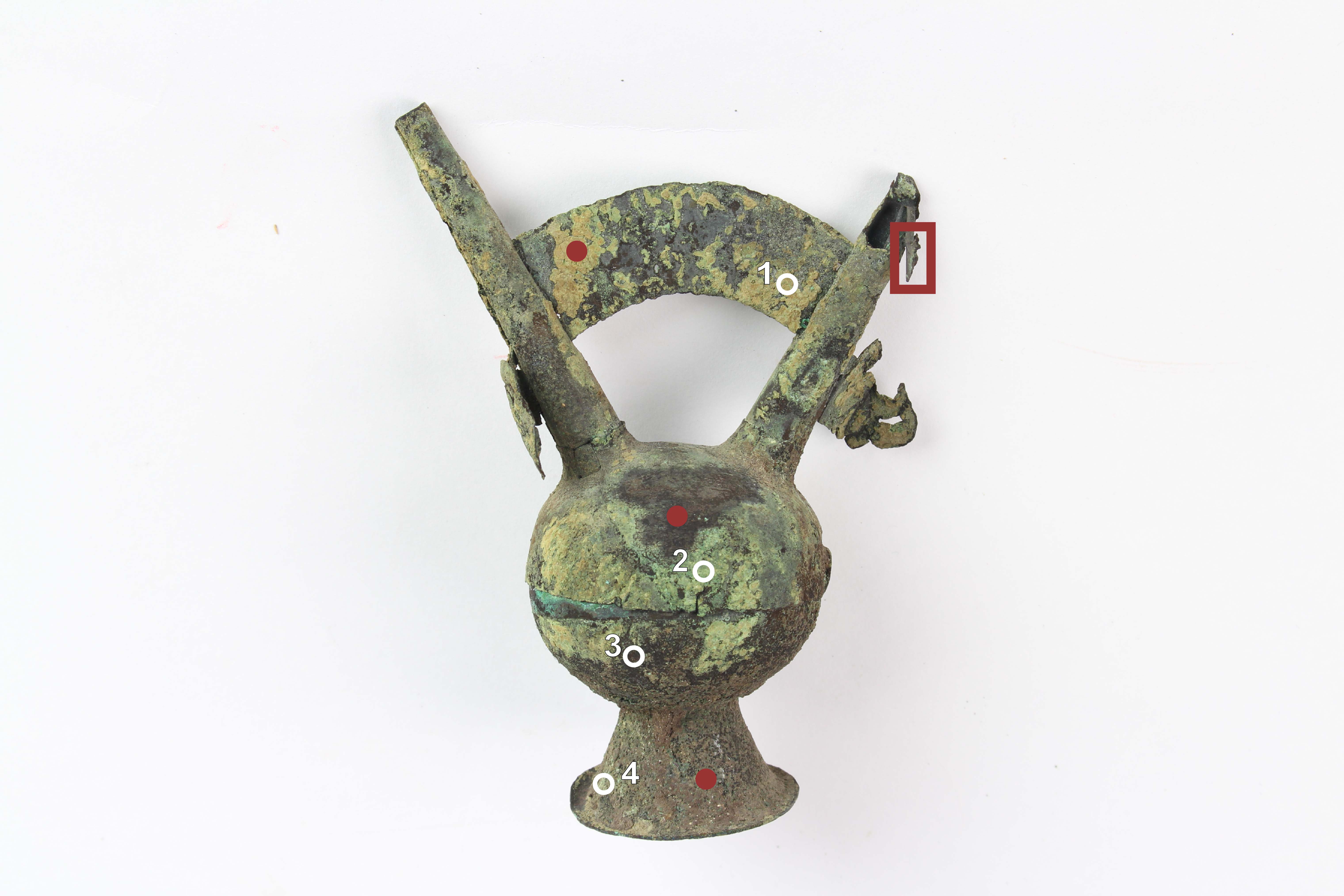
Peruvian funerary flask, approximately 9.5 cm high, which consists of two hemispheres joined at their centers. They are held at the sides by stape-like fasteners and attached to a hollow, flared base. Two long, hollow cylindrical tubes rise from the top of the sphere (Fig. 1).
Funeral flask
Lambayeque, Peru
Date of excavation unkown, acquisition in 1932
Medieval times
Could be from the Moche (100-700), Sicàn (700-1350) or Chimù (900-1450) civilization
Unknown
Kulturmuseum St-Gallen
Kulturmuseum St-Gallen
VK_D_1783
Ancient inventory informations
None.
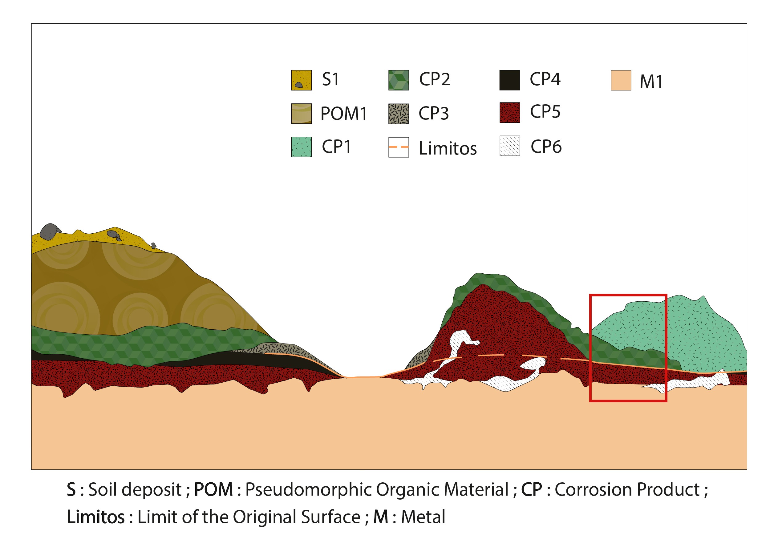
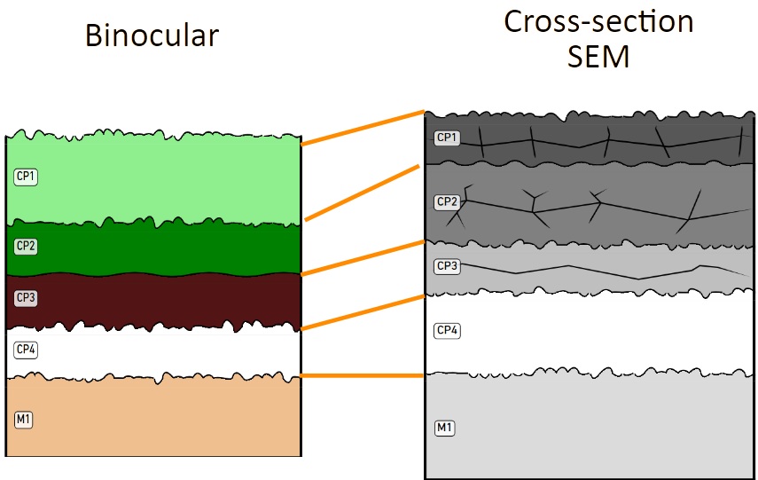
The schematic representation below gives an overview of the corrosion layers encountered on the funerary flask from the visual macroscopic observation.
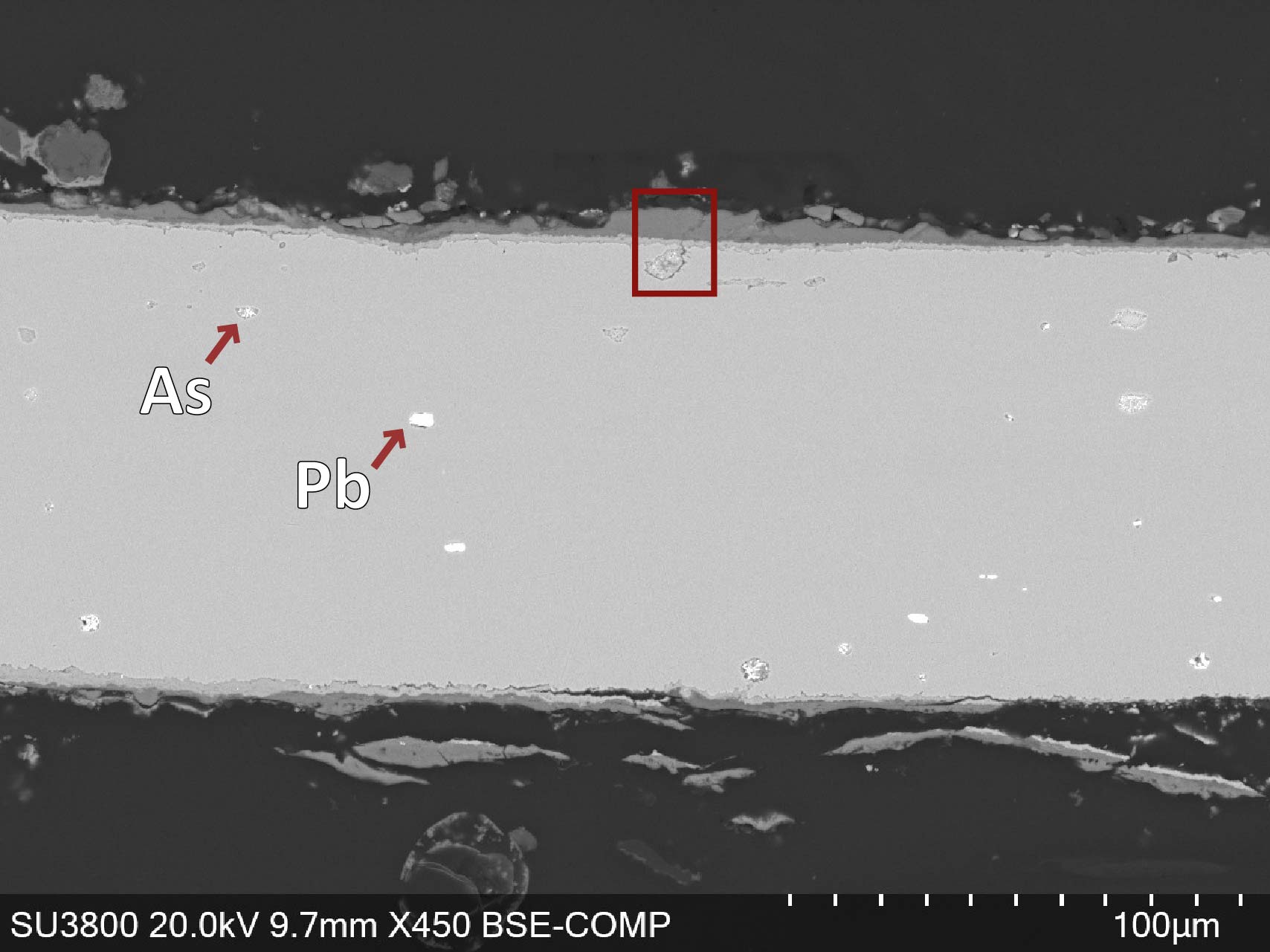
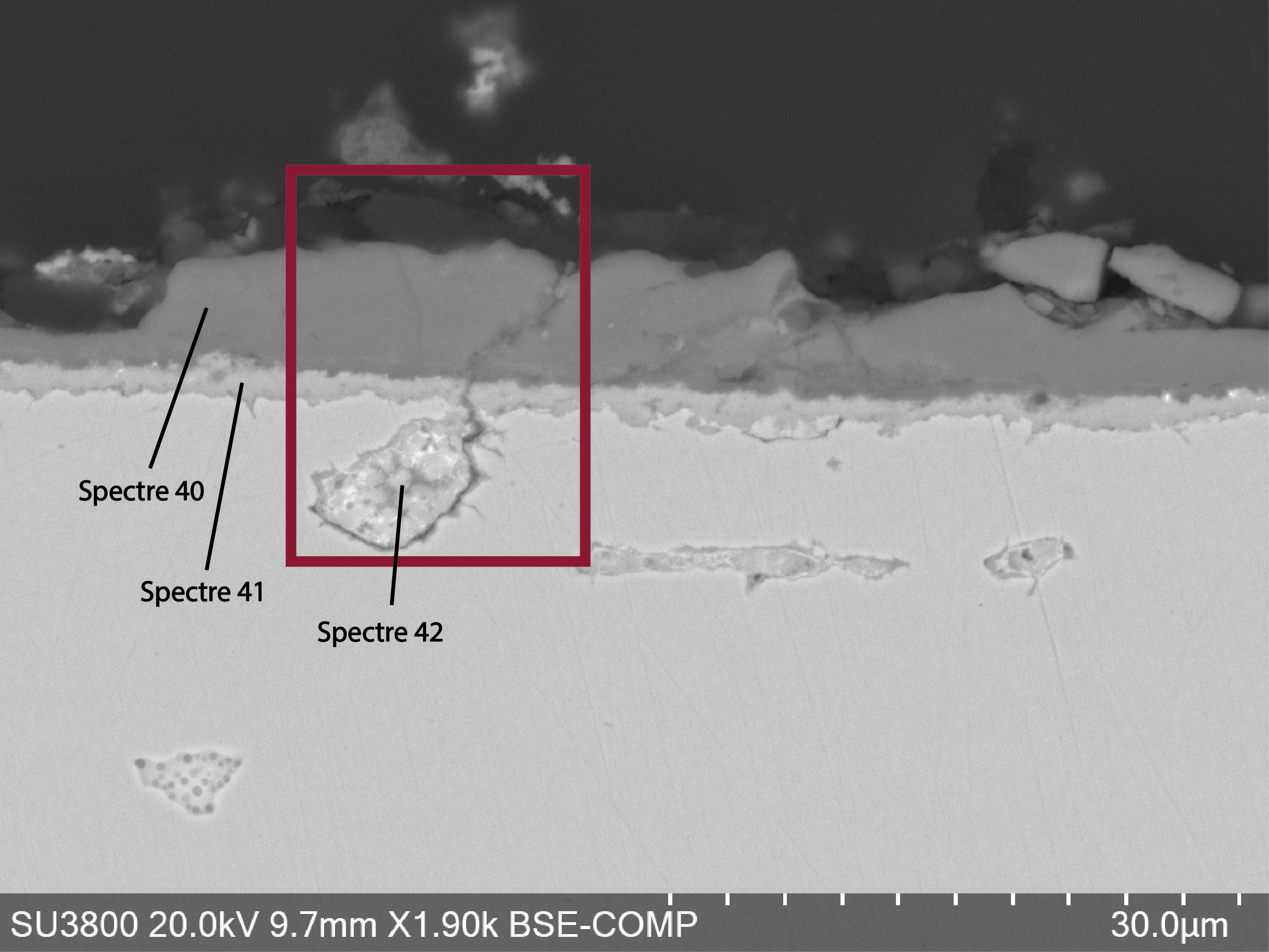
A polished cross-section was made from the already twisted damaged part on the flask (red square on Fig. 2) and embedded in resin. We can see the thin layer of corrosion mostly on the left side of the metal.
Cu/As/Pb
Hammered
None
Haute École Arc Neuchâtel, Neuchâtel
Kulturmuseum St-Gallen
2026, metallography and chemical analyses
None.
Analysis performed :
Non-invasive approach
- XRF, handheld device Thermo ScientificTM Nixton XL3t Goldd +, general metal mode (20s Main / 20s Low / 20 s Light) and mining mode (20s Main / 20s Lw / 20s High / 20s Light),
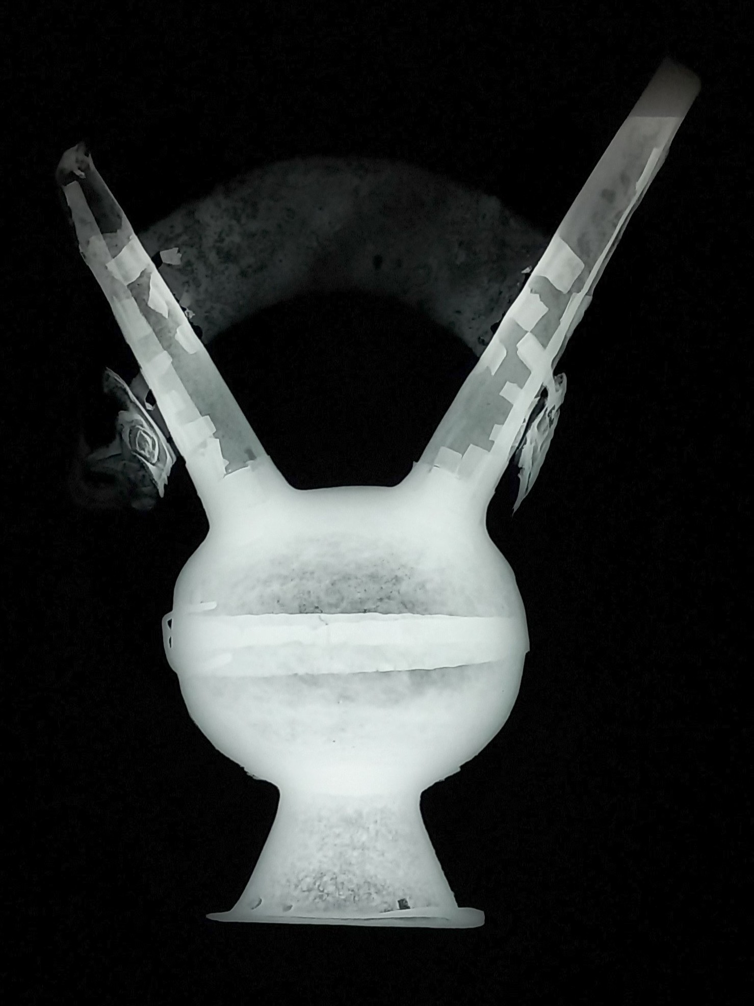
- X-radiography (44kV, 5 mA, 60s, d = 100cm).
Invasive approach
- SEM-EDS performed on individual samples and on the cross-section with an acceleration voltage of 20kV,
- Raman Spectroscopy (green laser).
The XRF analysis showed that the metal is probably a copper alloy, containing both lead (Pb) and Arsenic (As) in small concentrations (<1% mass).
X-ray radiography shows that the metal has been hammered repeatedly (Fig. 6).
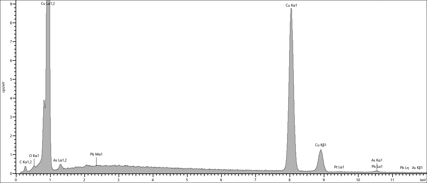
The metal consists of a copper-arsenic alloy (Table 1).
| Elements | Cu | As |
| Mass % | 98.8 | 1.2 |
Table 1: SEM-EDS analysis of the metal of the funerary flask (Fig. 7).
with lead and arsenic inclusions (Fig. 8).
The metal of this object is really thin and quite hard. Unfortunately the chemical etching that we tried on this cross-section did not give satisfactory results so we were not able to see the microstructure of the metal.
 Credit HEI-Arc, S.Ramseyer.
Unknown
Cu
As, Pb
None.
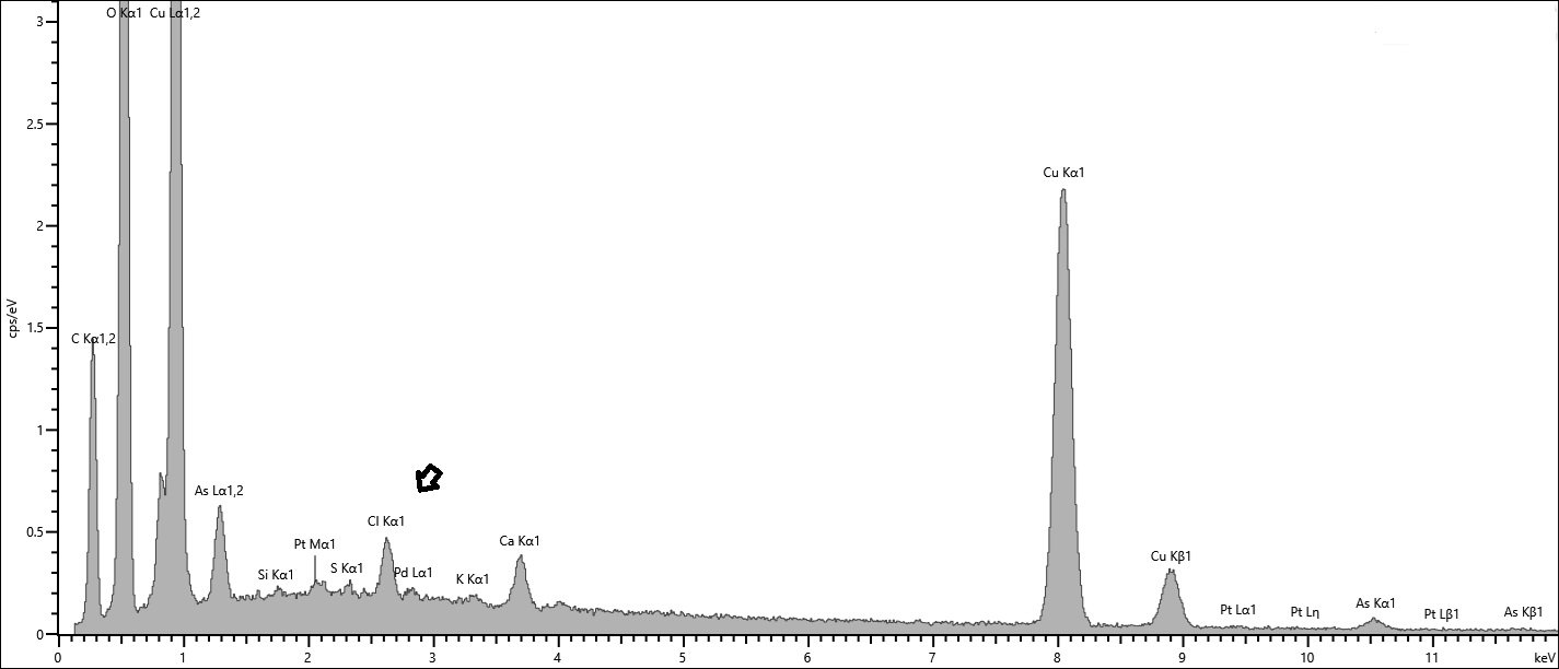
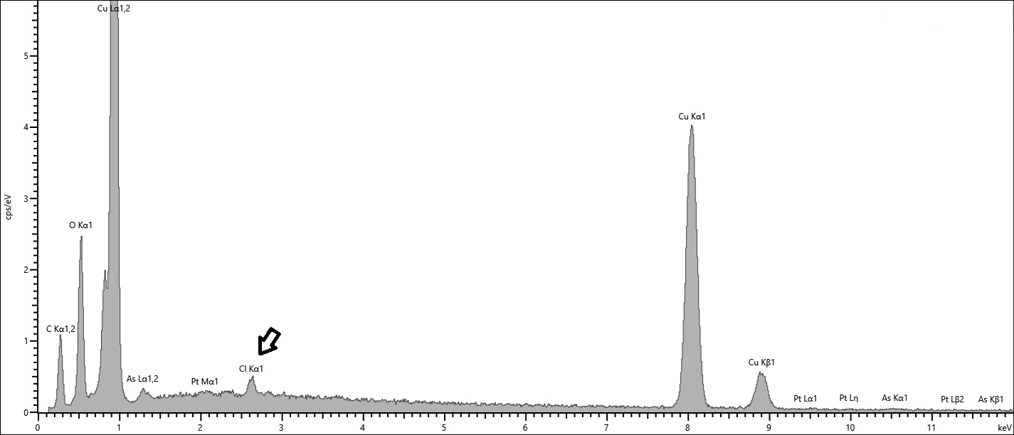
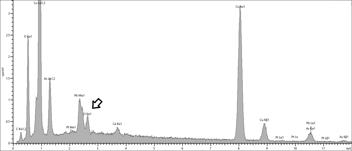
Even though active corrosion could not be clearly visualised optically, the examination of the cross-section revealed the presence of chlorides in the whole corrosion structure (Figures 9-13). These chlorides become active upon contact with oxygen and high relative humidity.
Active Corrosion (CP1 and CP6)
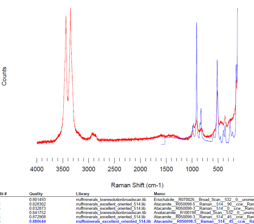
Chlorides from the environment entered the metal over centuries. They remain present throughout the metal and all corrosion layers. Upon excavation, exposure to oxygen and air humidity activated these chlorides. This activation is the root cause of the ongoing corrosion process. A white layer of nantokite (spectrum 42 - CP6, Fig. 9) was found near the sound metal and confirmed by EDS (Fig. 12), explaining the active corrosion. The powdery light green crystals on the top surface are atacamite (Fig. 9, CP1) a secondary corrosion product of nantokite. Its presence is confirmed by Raman spectroscopy (Fig. 13).
Mineralized Organic Remains (POM)
Circular micro-structures and a thicker corrosion layer identify mineralized organic remains on the vials. SEM analysis failed due to sample fragility during removal. Specialist examination ruled out textile and suggested mineralized leather instead. Traces appear on both faces, the sphere, and the transverse arch, indicating the object was likely entirely wrapped.
Black Corrosion (CP3 and CP4)
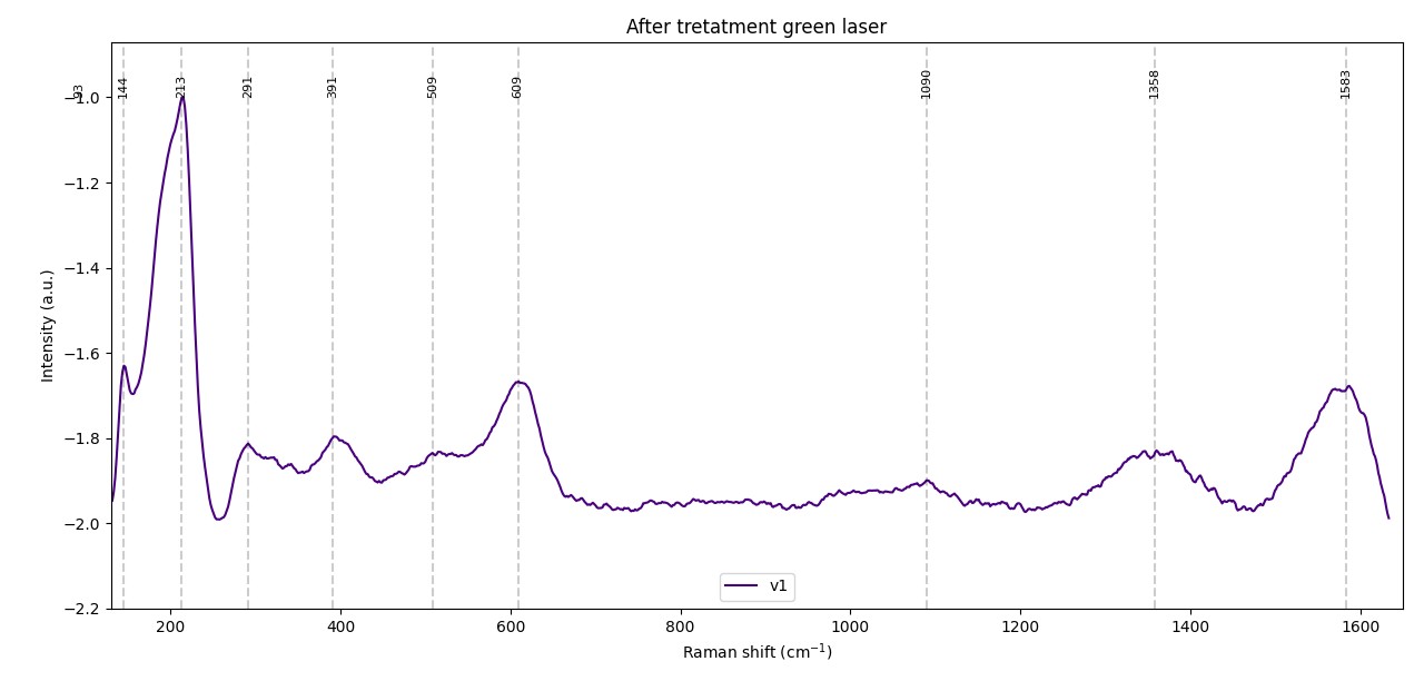
Black tufted corrosion from copper sulfides affects the flask on large areas. It likely originated from sulfate-reducing bacteria in anaerobic burial sediments. Raman analysis seems to indicate the presence of covellite (CuS, Fig. 14). This sulfide appears powdery on outer layers and crystalline where oxygen was limited.
Stable Corrosion and Metal State (CP2 and CP5)
Cuprite (CP5, Fig. 11) and malachite (?, CP2, Fig. 10) layers are stable, classic alteration products that protect the metal core. Unlike active corrosion, these layers are not deteriorating further. X-ray radiography confirmed the underlying metal remains sound. Gray variations on the X-ray image reflect the object's thickness and 3D shape, not metal degradation.
 Credit HEI-Arc, S.Ramseyer.
Multiform
Unknown
None.
The observation under binocular microscope and cross-section both identified 4 similar CPs with different widths though.
Using a binocular microscope, it is possible to distinguish the strata based on texture and color variations, whereas in cross-section (SEM), these strata exhibit different shades of gray, even though they are composed of similar elements at varying concentrations.
The metal is arsenical copper containing some lead inclusions. It appears to have been hammered repeatedly. The corrosion layers are typical of buried archaeological metal: cuprite, sulfides, and hydroxycarbonates. Chlorides have been detected and could trigger active corrosion.
References on object and analytical methods
- Scott, D. A. (1992) Metallography and microstructure in ancient and historic metals. Getty publications.
- Scott, D. A. (2002) Copper and bronze in art: corrosion, colorants, conservation. Getty publications.