Tang fragment of a knife HR-6567
Marianne. Senn (EMPA, Dübendorf, Zurich, Switzerland) & Christian. Degrigny (HE-Arc CR, Neuchâtel, Neuchâtel, Switzerland)
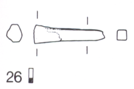
Tang fragment with shiny brown patina typical of lake context (Fig.1). Dimensions: L = 2.9cm; Ømax. = 6.8mm; WT = 4.9g.
Knife
Hauterive - Champréveyres, Neuchâtel, Neuchâtel, Switzerland
Excavation in 1983-1985, layer 3
Late Bronze Age
Hallstatt B1 (1054/1037BC _ 1000BC)
Lake
Laténium, Neuchâtel, Neuchâtel
Laténium, Neuchâtel, Neuchâtel
Hr 6567
Not conserved
Nothing to report.
Stratigraphic representation: none.
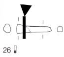
This cross-section shows a lateral cut through the tang (Fig. 2). Most of the corrosion crust is absent (Fig. 3).
Leaded Bronze
Cold worked after annealing
MAH 87-196
Musées d'art et d'histoire, Genève, Geneva
Musées d'art et d'histoire, Genève, Geneva
1987, metallography and corrosion characterisation
Nothing to report.
Analyses performed:
Metallography (etched with ferric chloride reagent), Vickers hardness testing, ICP-OES, SEM/EDS, XRD.
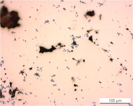
The remaining metal is a leaded bronze (Table 1) containing numerous copper sulphide and tiny Pb inclusions (Figs. 5-7, 10 and Table 2). The porosity within the metal is high, particularly along a band through the middle of the sample (Figs. 3 and 5). The etched structure of the leaded bronze shows small, regular polygonal grains, some with twinning (Fig. 6). Strain lines appear in grains close to the metal surface (Fig. 7). The average hardness of the metal is HV1 120.
| Elements | Cu | Sn | Pb | Ni | Sb | As | Co | Ag | Fe | Zn |
|---|---|---|---|---|---|---|---|---|---|---|
| mass% | 87.52 | 8.02 | 1.46 | 1.04 | 0.81 | 0.60 | 0.24 | 0.21 | 0.05 | 0.03 |
Table 1: Chemical composition of the metal. Method of analysis: ICP-OES, Laboratory of Analytical Chemistry, Empa.
| Elements | O | S | Fe | Cu | Total |
|---|---|---|---|---|---|
| mass% | 1.5 | 20 | 1.0 | 71 | 93 |
Table 2: Chemical composition of dark-grey inclusions. Method of analysis: SEM/EDS, Laboratory of Analytical Chemistry, Empa.
 Credit HE-Arc CR.
 Credit HE-Arc CR.
Polygonal and twinned grains + strain lines (metal surface) with pores
Cu
Co, Ni, As, Ag, Sn, Sb, Pb
Nothing to report.
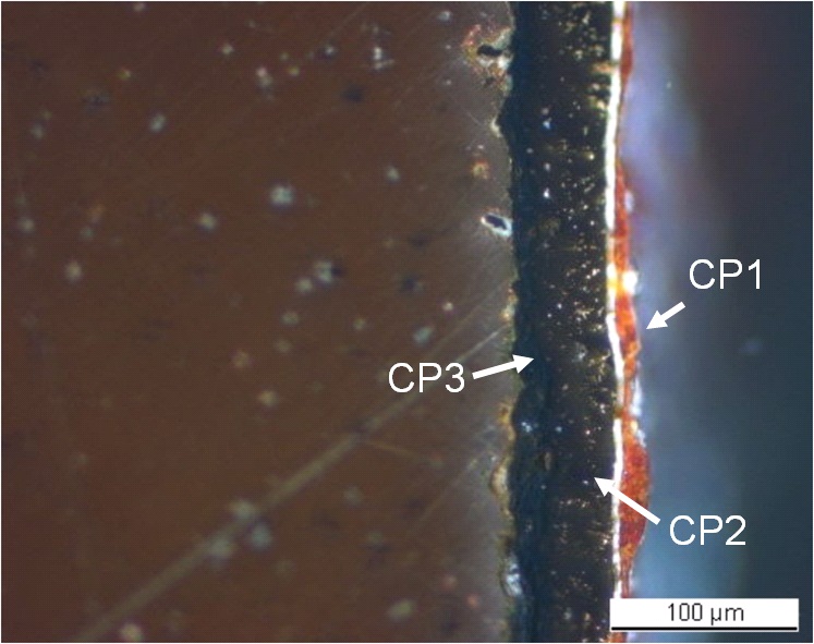
The metal has lost most of its original corrosion crust, the remainder having an average thickness between 60 and 190µm (Fig. 3). In some areas up to three corrosion layers are visible (Fig. 8). In polarised light (Fig. 9), the corrosion stratigraphy appears more clearly: it is composed of a dense black inner layer, an intermediate thick brown layer with bright spots (indicating porosity) and an outer red layer with white particles. The elemental chemical distribution of the SEM image reveals that the black inner layer (CP3) is Sn-rich, but contains Cu, O, Fe, Si, P, Pb, Ni, As, Ca and S (Table 3, Fig. 10). The brown layer (CP2) contains S, Fe and Cu and has a composition similar to chalcopyrite/CuFeS2 (Table 3, Fig. 10). This was confirmed by past XRD analyses carried out by Schweizer (1994, museum report (1987)). The red layer (CP1) is an iron oxide (main elements Fe and O) and is contaminated with calcite/CaCO3 particles (Table 3, Fig. 10).
|
Elements |
O | Fe | Ni | Cu | Si | P | S | Ca | As | Sn | Pb | Total |
|---|---|---|---|---|---|---|---|---|---|---|---|---|
| CP1, red layer | 37 | 51 | 1.8 | < | < | < | < | 1.5 | 0.8 | < | < | 93 |
| CP2, brown layer | < | 30 | < | 42 | < | < | 35 | < | < | < | < | 107 |
| CP2, white particles | 50 | < | < | 0.6 | < | < | < | 39 | < | < | < | 90 |
| CP3, black layer | 39 | 4.8 | 1.2 | 5.2 | 3.9 | 3.7 | < | < | 0.7 | 37 | 3.7 | 100 |
Table 3: Chemical composition (mass %) of the corrosion layers (from Figs. 8 and 9). Method of analysis: SEM/EDS, Laboratory of Analytical Chemistry, Empa.
 Credit HE-Arc CR.
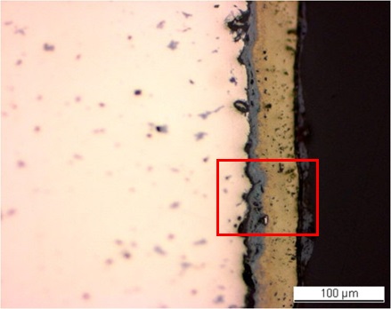
Fig. 8: Micrograph of the metal sample from Fig. 3 (reversed picture, detail), unetched, bright field. From left to right: metal (in pink), inner light-grey layer, intermediate brown layer and top dark-grey layer. The area selected for elemental chemical distribution (Fig. 10) is marked by a red rectangle,
 Credit HE-Arc CR.
Fig. 9: Micrograph of the same area as Fig. 8 and corresponding to the stratigraphy of Fig. 4, polarized light. From left to right: metal (in brown) covered with a corrosion crust consisting of a black layer, an intermediate brown layer with bright spots, a crack (white line) and a red layer with white particles,
Uniform - pitting
Type I (Robbiola)
Nothing to report.
Corrected stratigraphic representation: none.
The tang fragment is made from a leaded bronze and has been cold worked on the top surface after annealing. The SEM/EDS examination and past XRD analyses indicate the presence of chalcopyrite in the corrosion crust, typical of lake context (Schweizer 1994), enriched with Sn close to the metal surface and depleted of Cu on the outer surface. This object was certainly abandoned rather quickly in an anaerobic, humid and S and Fe-rich environment, favouring then the formation of chalcopyrite. The limit of the original surface most probably lies between the Sn-rich inner layer and the Fe and S-rich outer layers. The presence of iron oxides on top of the copper corrosion crust has not yet been explained. The corrosion is a type 1 according Robbiola et al. 1998.
|
References on object and sample |
|
References object References sample |
|
References on analytic methods and interpretation |
| 4. Robbiola, L., Blengino, J-M., Fiaud, C. (1998) Morphology and mechanisms of formation of natural patinas on archaeological Cu-Sn alloys, Corrosion Science, 40, 12, 2083-2111. |